Human MFAP3L Antibody
R&D Systems, part of Bio-Techne | Catalog # MAB11325

Key Product Details
Species Reactivity
Applications
Label
Antibody Source
Product Specifications
Immunogen
Met1-Met149
Accession # O75121
Specificity
Clonality
Host
Isotype
Scientific Data Images for Human MFAP3L Antibody
Detection of MFAP3L in RT-4 (Positive) & HL-60 (Negative).
MFAP3L was detected in immersion fixed RT-4 human urinary bladder transitional cell papilloma cell line (Positive) & absent in HL-60 human acute promyelocytic leukemia cell line (Negative) using Mouse Anti-Human MFAP3L Monoclonal Antibody (Catalog # MAB11325) at 8 µg/mL for 3 hours at room temperature. Cells were stained using the NorthernLights™ 557-conjugated Anti-Mouse IgG Secondary Antibody (red; Catalog # NL007) and counterstained with DAPI (blue). Specific staining was localized to cytoplasm. View our protocol for Fluorescent ICC Staining of Cells on Coverslips.Detection of MFAP3L in RT-4 (Positive) & THP-1 (Negative).
MFAP3L was detected in immersion fixed RT-4 human urinary bladder transitional cell papilloma cell line (Positive) & absent in THP-1 human acute monocytic leukemia cell line (Negative) using Mouse Anti-Human MFAP3L Monoclonal Antibody (Catalog # MAB11325) at 8 µg/mL for 3 hours at room temperature. Cells were stained using the NorthernLights™ 557-conjugated Anti-Mouse IgG Secondary Antibody (red; Catalog # NL007) and counterstained with DAPI (blue). Specific staining was localized to cytoplasm. View our protocol for Fluorescent ICC Staining of Cells on Coverslips.MFAP3L in SK-BR-3 (Positive) & HL-60 (Negative).
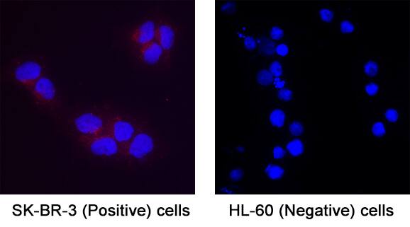
MFAP3L was detected in immersion fixed SK-BR-3 human breast cancer cell line (Positive) & absent in HL-60 human acute promyelocytic leukemia cell line (Negative) using Mouse Anti-Human MFAP3L Monoclonal Antibody (Catalog # MAB11325) at 8 µg/mL for 3 hours at room temperature. Cells were stained using the NorthernLights™ 557-conjugated Anti-Mouse IgG Secondary Antibody (red; Catalog # NL007) and counterstained with DAPI (blue). Specific staining was localized to cytoplasm. View our protocol for Fluorescent ICC Staining of Cells on Coverslips.Applications for Human MFAP3L Antibody
Immunocytochemistry
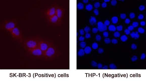
Sample: Immersion fixed RT‑4 human urinary bladder transitional cell papilloma cell line (Positive), SK‑BR‑3 human breast cancer cell line (Positive), HL‑60 human acute promyelocytic leukemia cell line (Negative) and THP‑1 human acute monocytic leukemia cell line (Negative).
Formulation, Preparation, and Storage
Purification
Reconstitution
Formulation
Shipping
Stability & Storage
- 12 months from date of receipt, -20 to -70 °C as supplied.
- 1 month, 2 to 8 °C under sterile conditions after reconstitution.
- 6 months, -20 to -70 °C under sterile conditions after reconstitution.
Background: MFAP3L
Microbibrillar-Associated Protein 3-Like (MFAP3L), also known as NYD-sp9, is part of the microfibrillar-associated protein family (MFAPs). MFAPs are non‐fibrillin, extracellular matrix glycoproteins that interact with fibrillin and were originally characterized in microfibrillar assembly (1,2). In humans, there several subfamily members with varying amino acid (aa) sequence homology and functions (1,2). Among the family, MFAP2 and MFAP5 are more closely related and while MFAP1, 3 and 4 share no structural or sequence homology with MFAP2, MFAP5 or with each other (1,2). Human MFAP3L shows 71% amino acid (aa) sequence homology to MFAP3, but not other MFAPs (3). Mature, human MFAP3L consists of an extracellular domain (ECD) containing N-linked glycosylation sites, a transmembrane domain, and a cytoplasmic domain with a conserved SH2 motif (3). The ECD of human MFAP3L shares 89% and 90% aa sequence identity with mouse and rat MFAP3L, respectively. MFAPs have the unique ability to interact with TGF-beta family growth factors, Notch and Notch ligands and multiple elastic fiber proteins, in addition to interacting with fibrillin (1, 2). MFAPs are expressed in a wide variety of tissues and, along with microfibril assembly, they play roles in the regulation of tissue homeostasis, cell survival, and tumor progression (1,2). MFAP3L is often located within colorectal cancer (CRC) cells, which metastasize by activation of the nuclear ERK pathway via MFAP3L phosphorylation (3). Regulation of this MFAP3L activity could have pharmaceutical effects on CRC tumor progression (3).
References
- Zhu, S. et al. (2020) J Cell Physio. 236:41.
- Mecham, R.P. et al. (2015) Matrix Biol. 47:13.
- Lou, X. et al. (2014) BBA. 1842:1423.
Long Name
Alternate Names
Gene Symbol
UniProt
Additional MFAP3L Products
Product Documents for Human MFAP3L Antibody
Product Specific Notices for Human MFAP3L Antibody
For research use only


