Human MICA Biotinylated Antibody
R&D Systems, part of Bio-Techne | Catalog # BAF1300


Key Product Details
Species Reactivity
Validated:
Cited:
Applications
Validated:
Cited:
Label
Antibody Source
Product Specifications
Immunogen
Ala22-Gln308
Accession # NP_001170990
Specificity
Clonality
Host
Isotype
Scientific Data Images for Human MICA Biotinylated Antibody
MICA in Human Colon.
MICA was detected in immersion fixed paraffin-embedded sections of human colon using Goat Anti-Human MICA Biotinylated Antigen Affinity-purified Polyclonal Antibody (Catalog # BAF1300) at 10 µg/mL overnight at 4 °C. Tissue was stained using the Anti-Goat HRP-DAB Cell & Tissue Staining Kit (brown; Catalog # CTS008) and counterstained with hematoxylin (blue). View our protocol for Chromogenic IHC Staining of Paraffin-embedded Tissue Sections.MICA in Human Colon.
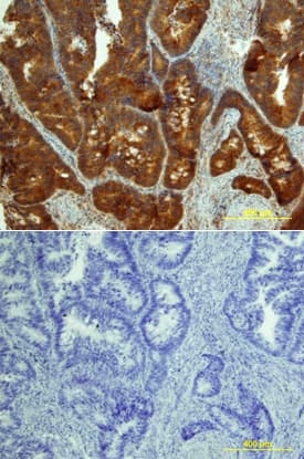
MICA was detected in immersion fixed paraffin-embedded sections of human colon array using Goat Anti-Human MICA Biotinylated Antigen Affinity-purified Polyclonal Antibody (Catalog # BAF1300) at 10 µg/mL overnight at 4 °C. Tissue was stained using the Anti-Goat HRP-DAB Cell & Tissue Staining Kit (brown; Catalog # CTS008) and counterstained with hematoxylin (blue). Lower panel shows a lack of labeling if primary antibodies are omitted and tissue is stained only with secondary antibody followed by incubation with detection reagents. View our protocol for Chromogenic IHC Staining of Paraffin-embedded Tissue Sections.Applications for Human MICA Biotinylated Antibody
Immunohistochemistry
Sample: Immersion fixed paraffin-embedded sections of human breast cancer tissue and human colon
Western Blot
Sample: Recombinant Human MICA Fc Chimera (Catalog # 1300-MA)
Human MICA Sandwich Immunoassay
Reviewed Applications
Read 1 review rated 3 using BAF1300 in the following applications:
Formulation, Preparation, and Storage
Purification
Reconstitution
Formulation
Shipping
Stability & Storage
- 12 months from date of receipt, -20 to -70 °C as supplied.
- 1 month, 2 to 8 °C under sterile conditions after reconstitution.
- 6 months, -20 to -70 °C under sterile conditions after reconstitution.
Background: MICA
MICA (MHC class I chain-related gene A) is a transmembrane glycoprotein that functions as a ligand for human NKG2D. A closely related protein, MICB, shares 85% amino acid identity with MICA. These proteins are distantly related to the MHC class I proteins. They possess three extracellular Ig-like domains, but they have no capacity to bind peptide or interact with beta2-microglobulin. The genes encoding these proteins are found within the Major Histocompatibility Complex on human chromosome 6. The MICA locus is highly polymorphic with more than 50 recognized human alleles. MICA is absent from most cells but is frequently expressed in epithelial tumors and can be induced by bacterial and viral infections. MICA is a ligand for human NKG2D, an activating receptor expressed on NK cells, NKT cells, gamma delta T cells, and CD8+ alpha beta T cells. Recognition of MICA by NKG2D results in the activation of cytolytic activity and/or cytokine production by these effector cells. MICA recognition is involved in tumor surveillance, viral infections, and autoimmune diseases.
References
- Groh, V. et al. (2001) Nature Immunol. 2:255.
- Stephens, H. (2001) Trends Immunol. 22:378.
- Bauer, S. et al. (1999) Science 285:727.
- Groh, V. et al. (2002) Nature 419:734.
- Steinle, A. et al. (2001) Immunogenetics 53:279.
- Pende, D. et al. (2002) Cancer Res. 62:6178.
- NKG2D and its Ligands (2002) http://www.RnDSystems.com.
Long Name
Alternate Names
Entrez Gene IDs
Gene Symbol
UniProt
Additional MICA Products
Product Documents for Human MICA Biotinylated Antibody
Product Specific Notices for Human MICA Biotinylated Antibody
For research use only
