Human MMP-7 Antibody
R&D Systems, part of Bio-Techne | Catalog # MAB9071


Key Product Details
Validated by
Species Reactivity
Validated:
Cited:
Applications
Validated:
Cited:
Label
Antibody Source
Product Specifications
Immunogen
Leu18-Lys267 (Ala230del)
Accession # NP_002414.1
Specificity
Clonality
Host
Isotype
Scientific Data Images for Human MMP-7 Antibody
Detection of Human MMP‑7 by Western Blot.
Western blot shows lysates of Capan-1 human pancreatic adenocarcinoma cell line and SK-OV-3 human ovarian adenocarcinoma cell line and, for additional reference, Recombinant Human MMP-7 Western Blot Standard Protein (Catalog # WBC016). PVDF membrane was probed with 1 µg/mL of Mouse Anti-Human MMP-7 Monoclonal Antibody (Catalog # MAB9071) followed by HRP-conjugated Anti-Mouse IgG Secondary Antibody (Catalog # HAF018). A specific band was detected for MMP-7 at approximately 28 kDa (as indicated). This experiment was conducted under reducing conditions and using Immunoblot Buffer Group 1.Detection of Human MMP‑7 by Simple WesternTM.
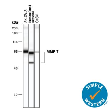
Simple Western lane view shows lysates of SK‑OV‑3 human ovarian adenocarcinoma cell line, human small intestine tissue, and CaSki human cervical carcinoma, loaded at 0.5 mg/mL. Specific bands were detected for MMP‑7 at approximately 49 and 65 kDa (as indicated) using 20 µg/mL of Mouse Anti-Human MMP-7 Monoclonal Antibody (Catalog # MAB9071) . This experiment was conducted under reducing conditions and using the 12-230 kDa separation system.Detection of Human MMP-7 by Western Blot
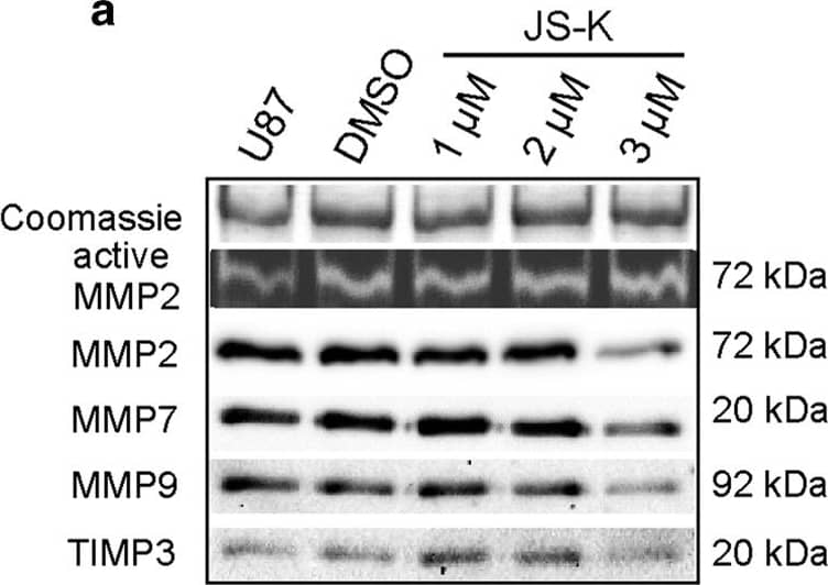
Zymography analysis for active MMP2 was performed with 4 μg of total protein of the conditioned medium of U87 (a) and primary IC cells (b) after 48 h exposure to JS-K (1–3 μM) on gelatin-containing PAGE. Western blot analysis for MMP2, MMP7, MMP9 and TIMP3 was performed for conditioned medium of U87 (a) and IC (b) cells with 4 μg of total protein as well as of 20 μg of total cell lysate of U87 (c) and IC cells (d) on SDS-PAGE. Conditioned media of knockdown and overexpression of ATF3 were examined for active MMP2 as well as the plasmid controls and uninfected U87 (e). 4 μg of protein of the supernatant (e) and 20 μg of cell lysates (f) were screened for MMP2, MMP7, MMP9 and TIMP3 by SDS-PAGE. Coomassie staining of the gels was used as a loading control for zymogram and for western blot of the supernatant, GAPDH was used as a loading control for cell lysates. Figures are representative for three independent experiments. Image collected and cropped by CiteAb from the following publication (https://pubmed.ncbi.nlm.nih.gov/28250971), licensed under a CC-BY license. Not internally tested by R&D Systems.Applications for Human MMP-7 Antibody
Immunohistochemistry
Sample: Immersion fixed paraffin-embedded sections of human pancreatic cancer tissue
Immunoprecipitation
Sample: Conditioned cell culture medium spiked with Recombinant Human MMP-7 (Catalog # 907-MP), see our available Western blot detection antibodies
Simple Western
Sample: SK‑OV‑3 human ovarian adenocarcinoma cell line, Human small intestine tissue, and CaSki human cervical carcinoma cell line
Western Blot
Sample: Capan‑1 human pancreatic adenocarcinoma cell line, SK‑OV‑3 human ovarian adenocarcinoma cell line, and Recombinant Human MMP-7 Western Blot Standard Protein (Catalog # WBC016).
Formulation, Preparation, and Storage
Purification
Reconstitution
Formulation
Shipping
Stability & Storage
- 12 months from date of receipt, -20 to -70 °C as supplied.
- 1 month, 2 to 8 °C under sterile conditions after reconstitution.
- 6 months, -20 to -70 °C under sterile conditions after reconstitution.
Background: MMP-7
Matrix metalloproteinases (MMPs) are a family of zinc and calcium dependent endopeptidases with the combined ability to degrade all the components of the extracellular matrix. MMP-7 (matrilysin) is expressed in epithelial cells of normal and diseased tissues, and is capable of digesting a large series of proteins of the extracellular matrix including collagen IV and X, gelatin, casein, laminin, aggrecan, entactin, elastin and versican. MMP-7 is implicated in the activation of other proteinases such as plasminogen, MMP-1, MMP-2, and MMP-9. In addition to its roles in connective tissue remodeling and cancer, MMP-7 also regulates intestinal alpha‑defensin activation in innate host defense, releases tumor necrosis factor-alpha in a model of herniated disc resorption, and cleaves FasL to generate a soluble form in a model of prostate involution. Structurally, MMP-7 is the smallest of the MMPs and consists of two domains: a pro-domain that is cleaved upon activation and a catalytic domain containing the zinc-binding site.
Long Name
Alternate Names
Gene Symbol
UniProt
Additional MMP-7 Products
Product Documents for Human MMP-7 Antibody
Product Specific Notices for Human MMP-7 Antibody
For research use only

