Human/Mouse ASAM Antibody
R&D Systems, part of Bio-Techne | Catalog # AF7356

Key Product Details
Species Reactivity
Applications
Label
Antibody Source
Product Specifications
Immunogen
Thr18-Ala234
Accession # Q8R373
Specificity
Clonality
Host
Isotype
Scientific Data Images for Human/Mouse ASAM Antibody
Detection of Mouse ASAM by Western Blot.
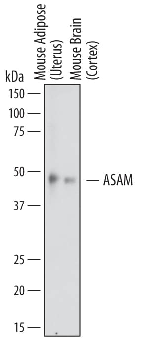
Western blot shows lysates of mouse adipose (uterus) tissue and mouse brain (cortex) tissue. PVDF membrane was probed with 1 µg/mL of Sheep Anti-Mouse ASAM Antigen Affinity-purified Polyclonal Antibody (Catalog # AF7356) followed by HRP-conjugated Anti-Sheep IgG Secondary Antibody (Catalog # HAF016). A specific band was detected for ASAM at approximately 48 kDa (as indicated). This experiment was conducted under reducing conditions and using Immunoblot Buffer Group 1.Detection of ASAM in RAW 264.7 Mouse Cell Line by Flow Cytometry.
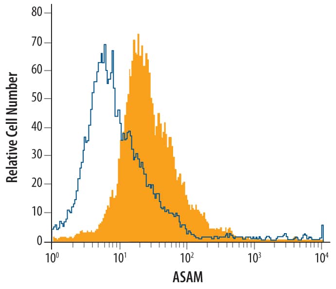
RAW 264.7 mouse monocyte/macrophage cell line was stained with Sheep Anti-Mouse ASAM Antigen Affinity-purified Polyclonal Antibody (Catalog # AF7356, filled histogram) or control antibody (Catalog # 5-001-A, open histogram), followed by Phycoerythrin-conjugated Anti-Sheep IgG Secondary Antibody (Catalog # F0126).Detection of ASAM in T98G Human Cell Line by Flow Cytometry.
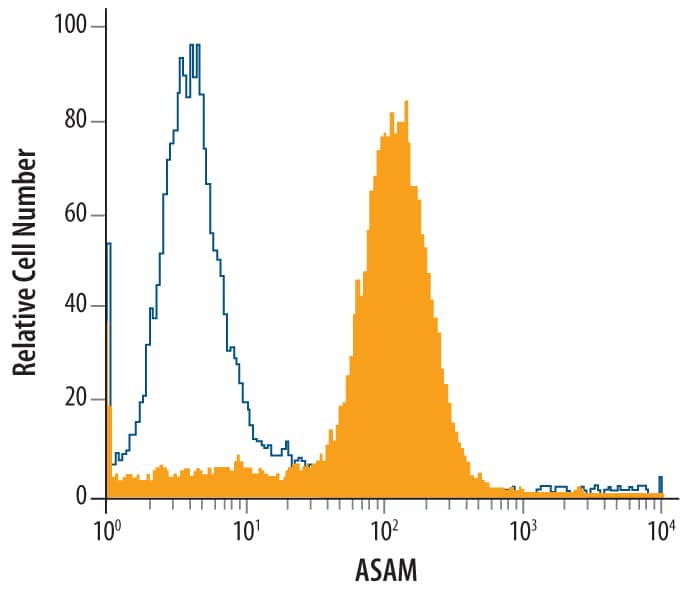
T98G human glioblastoma cell line was stained with Sheep Anti-Mouse ASAM Antigen Affinity-purified Polyclonal Antibody (Catalog # AF7356, filled histogram) or control antibody (Catalog # 5-001-A, open histogram), followed by Phycoerythrin-conjugated Anti-Sheep IgG Secondary Antibody (Catalog # F0126).Applications for Human/Mouse ASAM Antibody
CyTOF-ready
Flow Cytometry
Sample: T98G human glioblastoma cell line and RAW 264.7 mouse monocyte/macrophage cell line
Western Blot
Sample: Mouse adipose (uterus) tissue and mouse brain (cortex) tissue
Formulation, Preparation, and Storage
Purification
Reconstitution
Formulation
Shipping
Stability & Storage
- 12 months from date of receipt, -20 to -70 °C as supplied.
- 1 month, 2 to 8 °C under sterile conditions after reconstitution.
- 6 months, -20 to -70 °C under sterile conditions after reconstitution.
Background: ASAM
ASAM (Adipocyte-specific adhesion molecule; also ACAM, CLMP, Asp5 and CXADR-like protein) is a 44-48 kDa member of the CTX family of molecules. It is expressed on adipocytes as well as select epithelial cells such as respiratory and intestinal epithelium, keratinocytes and Sertoli cells. ASAM appears to contribute to tight junction formation, and may promote the transition of preadipocytes to adipocytes. Mature mouse ASAM is a 356 amino acid (aa) type I transmembrane glycoprotein. It contains a 217 aa extracellular region (aa 18-234) that is characterized by the presence of two C2 type Ig-like domains (aa 18-126 and 134-223), and possesses a C-terminal 118 aa cytoplasmic domain. The extracellular region of mouse ASAM (aa 18-234) shares 97% and 100% aa sequence identity with human and rat ASAM, respectively.
Long Name
Alternate Names
Gene Symbol
UniProt
Additional ASAM Products
Product Documents for Human/Mouse ASAM Antibody
Product Specific Notices for Human/Mouse ASAM Antibody
For research use only


