Human/Mouse CD109 PE-conjugated Antibody
R&D Systems, part of Bio-Techne | Catalog # FAB4385P


Key Product Details
Validated by
Species Reactivity
Validated:
Cited:
Applications
Validated:
Cited:
Label
Antibody Source
Product Specifications
Immunogen
Val22-Ser1268 (Tyr703Ser & Thr1241Met)
Accession # Q6YHK3
Specificity
Clonality
Host
Isotype
Scientific Data Images for Human/Mouse CD109 PE-conjugated Antibody
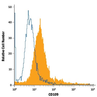
Detection of CD109 in A431 Human Cell Line by Flow Cytometry.
A431 human epithelial carcinoma cell line was stained with Mouse Anti-Human/Mouse CD109 PE-conjugated Monoclonal Antibody (Catalog # FAB4385P, filled histogram) or isotype control antibody (Catalog # IC003P, open histogram). View our protocol for Staining Membrane-associated Proteins.Detection of CD109 in Mouse Splenocytes by Flow Cytometry.
Mouse splenocytes treated with PMA and Calcium Ionomycin were stained with Mouse Anti-Human/Mouse CD109 PE-conjugated Monoclonal Antibody (Catalog # FAB4385P, filled histogram) or isotype control antibody (Catalog # IC003P, open histogram). View our protocol for Staining Membrane-associated Proteins.Detection of CD109 in Human PBMCs by Flow Cytometry.
Human peripheral blood mononuclear cells (PBMCs) treated with 2 ug/mL PHA for 5 days were stained with Mouse Anti-Human CD3e APC-conjugated Monoclonal Antibody (Catalog # FAB100A) and (A) Mouse Anti-Human CD109 PE-conjugated Monoclonal Antibody (Catalog # FAB4385P) or (B) PE-conjugated Mouse IgG2A isotype control (Catalog # IC003P). View our protocol for Staining Membrane-associated Proteins.Applications for Human/Mouse CD109 PE-conjugated Antibody
Flow Cytometry
Sample: A431 human epithelial carcinoma cell line, mouse splenocytes treated with PMA and Calcium Ionomycin, and human PBMC treated with PHA
Formulation, Preparation, and Storage
Purification
Formulation
Shipping
Stability & Storage
- 12 months from date of receipt, 2 to 8 °C as supplied.
Background: CD109
CD109 is a GPI-anchored member of the alpha-2-Macroglobulin (A2M) and complement family of proteins (1). Mature human CD109 contains a bait region with recognition sequences for multiple proteases, an internal thioester bond, and a domain similar to the receptor binding domain of A2M (2). Cleavage of A2M family proteins within the bait region activates the thioester bond to promote covalent bonding to nucleophilic groups in adjacent molecules (3, 4). Within the region included in this recombinant protein, human CD109 shares 71-73% amino acid (aa) sequence identity with mouse and rat CD109. It shares 27-33% aa sequence identity with A2M and complement factors C3, C4, and C5. Alternate splicing of human CD109 generates two isoforms with short deletions and one that is truncated within the bait region. CD109 is expressed on activated T cells and platelets, hematopoietic stem cells, megakaryocyte precursors, vascular endothelial cells, basal and myoepithelial cells of secretory glands, and squamous cell carcinomas (2, 5-9). It is produced as a 170-180 kDa glycoprotein that is autocatalytically processed to 150 kDa and 120 kDa forms (2, 6, 10). CD109 on keratinocytes binds TGF-beta and associates with TGF-beta RI and TGF-beta RII, resulting in inhibition of TGF-beta signaling (11). Polymorphisms of CD109 include the platelet-specific Gov antigen and the blood group ABH antigens (12, 13). Alloantibodies directed against these antigens result in unsuccessful platelet transfusions, neonatal alloimmune thrombocytopenia, and posttransfusion purpura (14).
References
- Travis, J. and G.S. Salvesen (1983) Annu. Rev. Biochem. 52:655.
- Lin, M. et al. (2002) Blood 99:1683.
- Christensen, U. and L. Sottrup-Jensen (1984) Biochemistry 23:6619.
- Wallis, R. et al. (2007) J. Biol. Chem. 282:7844.
- Murray, L.J. et al. (1999) Exp. Hematol. 27:1282.
- Haregewoin, A. et al. (1994) Cell. Immunol. 156:357.
- Hasegawa, M. et al. (2007) Pathol. Int. 57:245.
- Brashem-Stein, C. et al. (1988) J. Immunol. 140:2330.
- Hashimoto, M. et al. (2004) Oncogene 23:3716.
- Solomon, K.R. et al. (2004) Gene 327:171.
- Finnson, K.W. et al. (2006) FASEB J. 20:1525.
- Schuh, A.C. et al. (2002) Blood 99:1692.
- Kelton, J.G. et al. (1998) J. Lab. Clin. Med. 132:142.
- Rozman, P. (2002) Transpl. Immunol. 10:165.
Alternate Names
Gene Symbol
UniProt
Additional CD109 Products
Product Documents for Human/Mouse CD109 PE-conjugated Antibody
Product Specific Notices for Human/Mouse CD109 PE-conjugated Antibody
For research use only

