Human/Mouse DR3/TNFRSF25 Antibody
R&D Systems, part of Bio-Techne | Catalog # MAB943


Key Product Details
Species Reactivity
Validated:
Cited:
Applications
Validated:
Cited:
Label
Antibody Source
Product Specifications
Immunogen
Gln25-Phe201
Accession # Q93038
Specificity
Clonality
Host
Isotype
Scientific Data Images for Human/Mouse DR3/TNFRSF25 Antibody
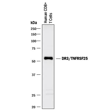
Detection of Human DR3/TNFRSF25 by Western Blot.
Western blot shows lysates of human CD8+T cells. PVDF membrane was probed with 2 µg/mL of Mouse Anti-Human/Mouse DR3/TNFRSF25 Monoclonal Antibody (Catalog # MAB943) followed by HRP-conjugated Anti-Mouse IgG Secondary Antibody (Catalog # HAF018). A specific band was detected for DR3/TNFRSF25 at approximately 55 kDa (as indicated). This experiment was conducted under reducing conditions and using Immunoblot Buffer Group 1.Applications for Human/Mouse DR3/TNFRSF25 Antibody
Western Blot
Sample: Human CD8+ T cells
Formulation, Preparation, and Storage
Purification
Reconstitution
Formulation
*Small pack size (-SP) is supplied either lyophilized or as a 0.2 µm filtered solution in PBS.
Shipping
Stability & Storage
- 12 months from date of receipt, -20 to -70 °C as supplied.
- 1 month, 2 to 8 °C under sterile conditions after reconstitution.
- 6 months, -20 to -70 °C under sterile conditions after reconstitution.
Background: DR3/TNFRSF25
Death receptor 3 (DR3), also known as lymphocyte-associated receptor of death (LARD), WSL-1, APO3, TRAMP and TR3, is a glycoprotein belonging to the TNF receptor superfamily (TNFRSF) (1 - 5). DR3 was formerly designated TNFRSF12 when it was thought to be a receptor for TWEAK/TNFSF12 (6). However, work disavowed the DR3:TWEAK interaction and DR3 is now designated TNFRSF 25 (7). By alternative splicing, at least 11 distinct human DR3 transcripts encoding secreted or type I membrane proteins exist (7). The human DR3 isoform 1 cDNA encodes a 417 amino acid residue (aa) transmembrane precursor with a 24 aa signal peptide, a 175 aa extracellular domain containing four cysteine-rich repeats and two potential N-glycosylation sites, a 21 aa transmembrane region and a 195 aa cytoplasmic region with one death domain. DR3 is one of six within the TNF R superfamily that contains a death domain in its cytooplasmic region. It is most closely related to TNF R1 and FAS/CD95, sharing 29% and 23% aa sequence identity, respectively. DR3 is expressed primarily in tissues enriched in lymphocytes. Whereas naïve B and T cells express multiple truncated DR3 isoforms but not the transmembrane isoform 1, upon T cell activation, expression of the transmembrane DR3 isoform 1 predominates. TL1A/VEGI, a TNF superfamily ligand, has been shown to bind and activate DR3 (8). Depending on the cell context, ligation of DR3 by TL1A can trigger one of two signaling pathways. On primary T cells, TL1A induces NF-kappa-B activation and a costimulatory signal to increase IL-2 responsiveness and the secretion of proinflammatory cytokines. However, in a tumor cell line, TF-1, TL1A has been shown to induce caspase activity and apoptosis. In DR3-null mice, an impairment of negative selection and anti-CD3-mediated thymocyte apoptosis is observed.
References
- Chinnaiyan, A.M. et al. (1996) Science 274:990.
- Kitson, J. et al. (1996) Nature 384:372.
- Bodmer, J-L. et al. (1997) Immunity 6:79.
- Screaton, G.R. et al. (1997) Proc. Nat. Acad. Sci. USA 94:4615.
- Marsters, S.A. et al. (1996) Curr. Biol. 6:1669.
- Marsters, S.A. et al. (1998) Curr. Biol. 8:525.
- Kaptein, A. et al. (2000) FEBS Lett. 485:135.
- Migone, T-S. et al. (2002) Immunity 16:479.
Long Name
Alternate Names
Gene Symbol
UniProt
Additional DR3/TNFRSF25 Products
Product Documents for Human/Mouse DR3/TNFRSF25 Antibody
Product Specific Notices for Human/Mouse DR3/TNFRSF25 Antibody
For research use only