Human/Mouse EBF-1 Antibody
R&D Systems, part of Bio-Techne | Catalog # AF5165

Key Product Details
Validated by
Species Reactivity
Validated:
Cited:
Applications
Validated:
Cited:
Label
Antibody Source
Product Specifications
Immunogen
Arg416-Ser520
Accession # Q07802
Specificity
Clonality
Host
Isotype
Scientific Data Images for Human/Mouse EBF-1 Antibody
Detection of Human/Mouse EBF-1 by Western Blot.
Western blot shows lysates of Raji human Burkitt's lymphoma cell line, Daudi human Burkitt's lymphoma cell line, and CH-1 mouse B cell lymphoma cell line, and L1.2 mouse pro-B cell line. PVDF membrane was probed with 2 µg/mL of Human/Mouse EBF-1 Antigen Affinity-purified Polyclonal Antibody (Catalog # AF5165) followed by HRP-conjugated Anti-Goat IgG Secondary Antibody (Catalog # HAF109). A specific band was detected for EBF-1 at approximately 70 kDa (as indicated). This experiment was conducted under reducing conditions and using Immunoblot Buffer Group 1.Detection of Human EBF-1 by Western Blot
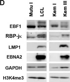
Differential binding of transcription factors on EBV genomes in type I and type III latency.(A) ChIP-Seq tracks mapped to EBV genome in LCL (red) or Mutu I (blue) for EBF1 or RBP-j kappa as indicated. EBNA2 ChIP-Seq for LCL (black). Y-axis represent raw read counts. (B) ChIP-qPCR for EBF1 or RBP-j kappa in LCL (red) or Mutu I (blue) at various EBV genome regulatory regions as indicated and Actin genomic region as negative control. (C) Same as in B, except for isogenic EBV positive BL cells Kem III (red) or Kem I (blue). Asterisk indicates p < 0.05. (D) Western blot of Mutu I, LCL, Kem I, and Kem III probed with antibody to EBF1, RBP-j kappa, LMP1, and EBNA2, with cellular loading controls for GAPDH and H3K4me3, as indicated. (E) RT-qPCR for EBV type III gene expression in LCL or Mutu I (left panel) or Kem I and Kem III (right panel). Image collected and cropped by CiteAb from the following publication (https://pubmed.ncbi.nlm.nih.gov/26752713), licensed under a CC-BY license. Not internally tested by R&D Systems.Detection of Human EBF-1 by Western Blot
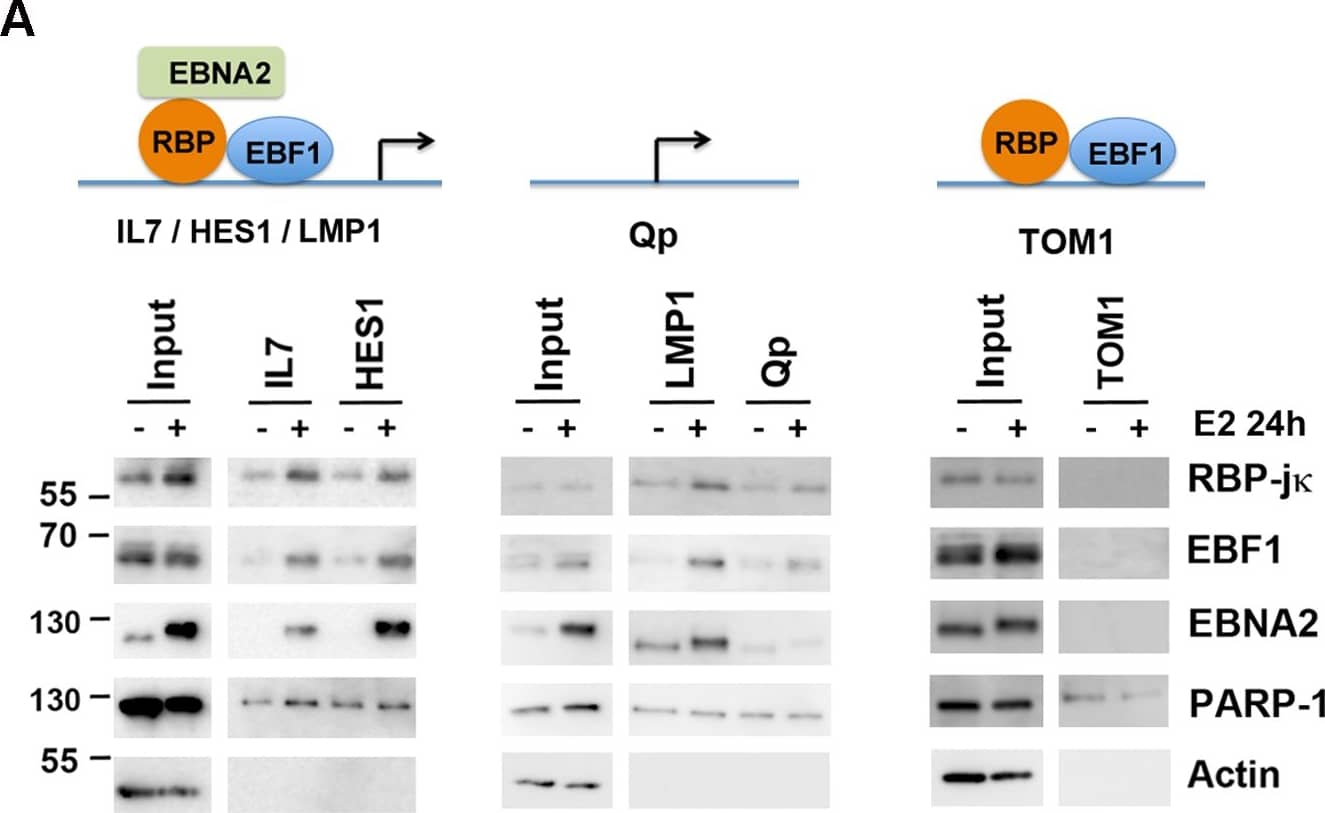
Cooperative binding and colocalization of EBF1, RBP-j kappa, and EBNA2 at DNA regulatory elements.(A) DNA-affinity binding assays with nuclear extracts from EREB2.5 cells with (+) or without (-) estradiol (E2) using DNA regulatory elements from IL7, HES1 (left), or LMP1, Qp (middle), or TOM1 (right). Input (10%) is indicated, and bound proteins were assayed by Western blot for EBF1, RBP-j kappa, EBNA2, and specificity controls for PARP1 and Actin. (B) ChIP-reChIP assays in LCLs with EBF1 as first ChIP, followed by a reChIP with either no antibody control or antibody to EBF1, RBP-j kappa or EBNA2. ReChIP DNA was quantified by qPCR at LMP1, IL7, and BDH2 (an EBF1 only site) binding sites. (C) Same as in B, except first ChIP was with RBP-j kappa followed by a ReChIP with no antibody control, or EBF1, RBP-j kappa, or EBNA2 antibody and assayed at LMP1, IL7, or LZTFL1 (an RBP-j kappa only site) binding sites. Asterisk indicates p < 0.05. Image collected and cropped by CiteAb from the following publication (https://pubmed.ncbi.nlm.nih.gov/26752713), licensed under a CC-BY license. Not internally tested by R&D Systems.Applications for Human/Mouse EBF-1 Antibody
Western Blot
Sample: Raji human Burkitt's lymphoma cell line, Daudi human Burkitt's lymphoma cell line, and CH-1 mouse B cell lymphoma cell line, and L1.2 mouse pro-B cell line
Formulation, Preparation, and Storage
Purification
Reconstitution
Formulation
Shipping
Stability & Storage
- 12 months from date of receipt, -20 to -70 °C as supplied.
- 1 month, 2 to 8 °C under sterile conditions after reconstitution.
- 6 months, -20 to -70 °C under sterile conditions after reconstitution.
Background: EBF-1
EBF-1 (Early B cell Factor 1; also OLF1 and COE1) is a 65-70 kDa member of the COE family of transcription factors. Although expressed in adipocytes and neurons, it is best studied in B cells where IL-7 acts to promote EBF-1 in pre-proB cells, leading to proB stage development. Mouse EBF-1 is 591 amino acids (aa) in length. It contains one DNA-binding region with an embedded zinc-finger motif (aa 51‑235), a dimerization segment between aa 370‑430, and a Pro/Ser-rich transactivation domain (aa 462‑550). EBF-1 either homodimerizes, or heterodimerizes with EBF-2 and -3. There is an alternate start site at Met134, and an isoform that shows a one aa substitution for aa 252‑259. Over aa 416‑520, mouse EBF-1 shows absolute aa identity to the equivalent sequence in rat and human EBF-1.
Long Name
Alternate Names
Gene Symbol
UniProt
Additional EBF-1 Products
Product Documents for Human/Mouse EBF-1 Antibody
Product Specific Notices for Human/Mouse EBF-1 Antibody
For research use only


