Human/Mouse Nogo-B Antibody
R&D Systems, part of Bio-Techne | Catalog # AF6034

Key Product Details
Species Reactivity
Applications
Label
Antibody Source
Product Specifications
Immunogen
Met1-Val200
Accession # NP_722550
Specificity
Clonality
Host
Isotype
Scientific Data Images for Human/Mouse Nogo-B Antibody
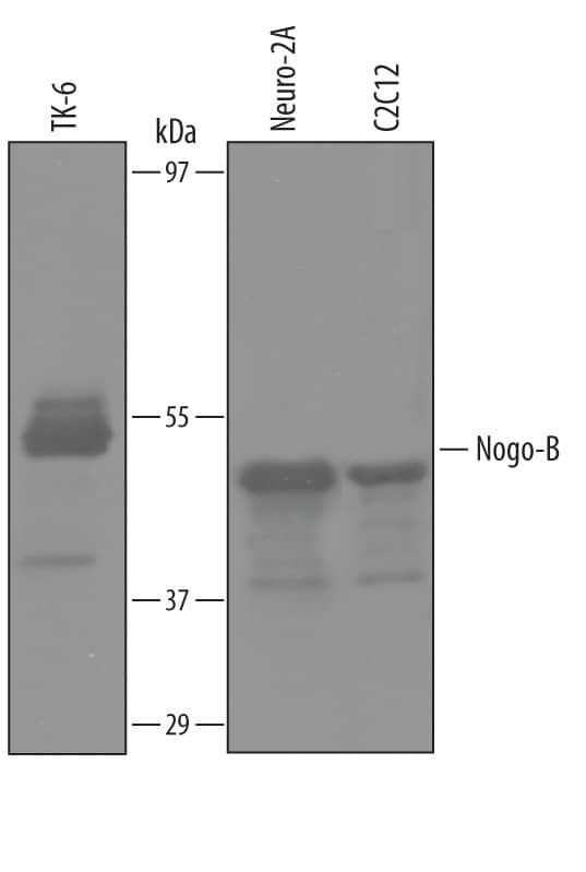
Detection of Human and Mouse Nogo‑B by Western Blot.
Western blot shows lysates of TK-6 human lymphoblast cell line, Neuro-2A mouse neuroblastoma cell line, and C2C12 mouse myoblast cell line. PVDF Membrane was probed with 1 µg/mL of Human/Mouse Nogo-B Antigen Affinity-purified Polyclonal Antibody (Catalog # AF6034) followed by HRP-conjugated Anti-Sheep IgG Secondary Antibody (HAF016). A specific band was detected for Nogo-B at approximately 49 kDa (as indicated). This experiment was conducted under reducing conditions and using Immunoblot Buffer Group 8.Nogo-B in Human Heart.
Nogo-B was detected in immersion fixed paraffin-embedded sections of human heart using Human/Mouse Nogo-B Antigen Affinity-purified Polyclonal Antibody (Catalog # AF6034) at 3 µg/mL overnight at 4 °C. Before incubation with the primary antibody, tissue was subjected to heat-induced epitope retrieval using Antigen Retrieval Reagent-Basic (CTS013). Tissue was stained using the Anti-Sheep HRP-DAB Cell & Tissue Staining Kit (brown; CTS019) and counterstained with hematoxylin (blue). Specific staining was localized to endothelial cells. View our protocol for Chromogenic IHC Staining of Paraffin-embedded Tissue Sections.Applications for Human/Mouse Nogo-B Antibody
Immunohistochemistry
Sample: Immersion fixed paraffin-embedded sections of human heart
Western Blot
Sample: TK‑6 human lymphoblast cell line, Neuro‑2A mouse neuroblastoma cell line, and C2C12 mouse myoblast cell line
Formulation, Preparation, and Storage
Purification
Reconstitution
Formulation
Shipping
Stability & Storage
- 12 months from date of receipt, -20 to -70 °C as supplied.
- 1 month, 2 to 8 °C under sterile conditions after reconstitution.
- 6 months, -20 to -70 °C under sterile conditions after reconstitution.
Background: Nogo-B
Nogo-B (A No-Go for neurite outgrowth Isoform B; also reticulon-4 isoform 2 and RTN-xS) is a 49‑51 kDa member of the reticulon protein family. It is widely expressed, being reported everywhere but liver. Nogo-B appears to be principally expressed in the ER, but does have a receptor (NgBR) on cell surfaces. Intracellularly, Nogo-B is known to interact with Bcl-xl and Bcl-2, and may play in role in both apoptosis and angiogenesis. Human Nogo-B is a two transmembrane, 373 amino acid protein. It contains an N-terminal cytoplasmic domain (aa 1‑199), two transmembrane segments (aa 200‑219 and 315‑335), a luminal region (aa 220‑314) and a C-terminal cytoplasmic domain (aa 336‑373). Nogo-B contains multiple phosphorylation sites and undergoes acetylation. Caspase-7 cleaves Nogo‑B between Asp14Ser15, generating a 45‑47 kDa fragment. Nogo-B exists in two forms; B1 represents aa 1‑185 spliced to aa 1005‑1192 of Nogo-A, while B2 represents aa 1‑204 spliced to aa 1005‑1192 of Nogo-A. Over aa 1‑200 (of Nogo-A), human Nogo-B shares 76% aa identity with mouse Nogo-B.
Long Name
Alternate Names
Gene Symbol
UniProt
Additional Nogo-B Products
Product Documents for Human/Mouse Nogo-B Antibody
Product Specific Notices for Human/Mouse Nogo-B Antibody
For research use only

