Human/Mouse/Rat Acetyl-CoA
Carboxylase alpha/ACACA Antibody
R&D Systems, part of Bio-Techne | Catalog # AF6898

Key Product Details
Species Reactivity
Applications
Label
Antibody Source
Product Specifications
Immunogen
Pro1185-Phe1352
Accession # Q13085
Specificity
Clonality
Host
Isotype
Scientific Data Images for Human/Mouse/Rat Acetyl-CoACarboxylase alpha/ACACA Antibody
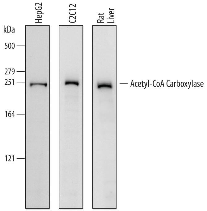
Detection of Human, Mouse, and Rat Acetyl-CoA Carboxylase alpha/ACACA by Western Blot.
Western blot shows lysates of HepG2 human hepatocellular carcinoma cell line, C2C12 mouse myoblast cell line, and rat liver tissue. PVDF membrane was probed with 0.5 µg/mL of Sheep Anti-Human/Mouse/Rat Acetyl-CoA Carboxylase a/ACACA Antigen Affinity-purified Polyclonal Antibody (Catalog # AF6898) followed by HRP-conjugated Anti-Sheep IgG Secondary Antibody (HAF016). A specific band was detected for Acetyl-CoA Carboxylase a/ACACA at approximately 250 kDa (as indicated). This experiment was conducted under reducing conditions and using Immunoblot Buffer Group 1.Detection of Human/Mouse/Rat Acetyl-CoA Carboxylase alpha/ACACA by Simple WesternTM.
Simple Western lane view shows lysates of A431 human epithelial carcinoma cells and C2C12 mouse myoblast cells, loaded at 0.2 mg/mL. A specific band was detected for Acetyl-CoA Carboxylase alpha/ACACA at approximately 380-400 kDa (as indicated) using 20 µg/mL of Sheep Anti-Human/Mouse/Rat Acetyl-CoA Carboxylase alpha/ACACA Antigen Affinity-purified Polyclonal Antibody (Catalog # AF6898) followed by 1:50 dilution of HRP-conjugated Anti-Sheep IgG Secondary Antibody (Catalog # HAF016). This experiment was conducted under reducing conditions and using the 66-440 kDa separation system.Applications for Human/Mouse/Rat Acetyl-CoACarboxylase alpha/ACACA Antibody
Simple Western
Sample: A431 human epithelial carcinoma cells and C2C12 mouse myoblast cells
Western Blot
Sample: HepG2 human hepatocellular carcinoma cell line, C2C12 mouse myoblast cell line, and rat liver tissue
Formulation, Preparation, and Storage
Purification
Reconstitution
Formulation
Shipping
Stability & Storage
- 12 months from date of receipt, -20 to -70 °C as supplied.
- 1 month, 2 to 8 °C under sterile conditions after reconstitution.
- 6 months, -20 to -70 °C under sterile conditions after reconstitution.
Background: Acetyl-CoA Carboxylase alpha/ACACA
ACAC-A (Acetyl-CoA carboxylase alpha/1; also ACC-1 and biotin carboxylase) is a 260-265 kDa cytoplasmic, phosphorylated biotinyl-enzyme. It is widely expressed, and found to be concentrated in hepatocytes, adipocytes and lactating mammary epithelium. It is one of two gene products (ACAC-B/beta being the other) that catalyze the formation of malonyl-CoA from acetyl-CoA. The formation of malonyl-CoA by ACAC-A is a rate-limiting step in fatty acid synthesis; malonyl-CoA formed by ACAC-B acts as a regulator of CPT-1 during fatty acidoxidation. Human ACAC-A is 2346 amino acids (aa ) in length. It contains an N-terminal acetylated Met, one ATP-Grasp domain (aa 275-466) with an embedded biotin carboxylation domain (aa 117-618), a biotinyl-binding region (aa 752-818), and a carboxyltransferase domain (aa 1698-2194). There are at least 17 utilized phosphorylation sites, and two acetylated Lys. ACAC-A exists as either a dimer or higher-order oligomer. Multiple splice variants exist. One possesses an alternative start site at Met79, a second utilizes an alternative start site 37 aa upstream of the standard site, and a third (called PIII) shows a 17 aa substitution for aa 1-75. Over aa 1185-1352, human ACAC-A shares 95% aa identity with mouse ACAC-A, and 97% aa identity with both ovine and bovine ACAC-A.
Long Name
Alternate Names
Gene Symbol
UniProt
Additional Acetyl-CoA Carboxylase alpha/ACACA Products
Product Documents for Human/Mouse/Rat Acetyl-CoACarboxylase alpha/ACACA Antibody
Product Specific Notices for Human/Mouse/Rat Acetyl-CoACarboxylase alpha/ACACA Antibody
For research use only

