Human/Mouse/Rat APE Antibody
R&D Systems, part of Bio-Techne | Catalog # AF1044

Key Product Details
Species Reactivity
Validated:
Cited:
Applications
Validated:
Cited:
Label
Antibody Source
Product Specifications
Immunogen
Pro2-Leu318
Accession # P27695
Specificity
Clonality
Host
Isotype
Scientific Data Images for Human/Mouse/Rat APE Antibody
Detection of Human, Mouse, and Rat APE by Western Blot.
Western blot shows lysates of HepG2 human hepatocellular carcinoma cell line, Balb/3T3 mouse embryonic fibroblast cell line, and Rat-2 rat embryonic fibroblast cell line. PVDF membrane was probed with 1 µg/mL of Goat Anti-Human/Mouse/Rat APE Antigen Affinity-purified Polyclonal Antibody (Catalog # AF1044) followed by HRP-conjugated Anti-Goat IgG Secondary Antibody (HAF017). A specific band was detected for APE at approximately 40 kDa (as indicated). This experiment was conducted using Immunoblot Buffer Group 1.Detection of Human APE by Simple WesternTM.
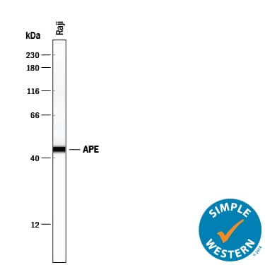
Simple Western lane view shows lysates of Raji human Burkitt's lymphoma cell line, loaded at 0.2 mg/mL. A specific band was detected for APE at approximately 45 kDa (as indicated) using 10 µg/mL of Goat Anti-Human/Mouse/Rat APE Antigen Affinity-purified Polyclonal Antibody (Catalog # AF1044) followed by 1:50 dilution of HRP-conjugated Anti-Goat IgG Secondary Antibody (HAF109). This experiment was conducted under reducing conditions and using the 12-230 kDa separation system.Detection of APE in A549 human lung carcinoma cells.
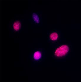
APE was detected in immersion fixed A549 human lung carcinoma cells using Goat Anti-Human/Mouse/Rat APE Antigen Affinity-purified Polyclonal Antibody (Catalog # AF1044) at 5 µg/mL for 3 hours at room temperature. Cells were stained using the NorthernLights™ 557-conjugated Anti-Goat IgG Secondary Antibody (red; Catalog # NL001) and counterstained with DAPI (blue). Specific staining was localized to Nuclear. View our protocol for Fluorescent ICC Staining of Cells on Coverslips.Applications for Human/Mouse/Rat APE Antibody
Immunocytochemistry
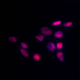
Sample: Immersion fixed A549 human lung carcinoma cells (Positive) and HepG2 human hepatocellular carcinoma cells (Positive)
Simple Western
Sample: Raji human Burkitt's lymphoma cell line
Western Blot
Sample: HepG2 human hepatocellular carcinoma cell line, Balb/3T3 mouse embryonic fibroblast cell line, and Rat-2 rat embryonic fibroblast cell line
Formulation, Preparation, and Storage
Purification
Reconstitution
Formulation
Shipping
Stability & Storage
- 12 months from date of receipt, -20 to -70 °C as supplied.
- 1 month, 2 to 8 °C under sterile conditions after reconstitution.
- 6 months, -20 to -70 °C under sterile conditions after reconstitution.
Background: APE
Human APE (also known as Ref-1) is the apurinic/apyrimidinic (AP) endonuclease required for efficient DNA base excision repair (BER). Following the removal of a damaged base by a DNA glycosylase, APE cleaves the AP site to allow resynthesis and ligation to complete repair. In addition, APE/Ref-1 acts as a factor that regulates the redox state of multiple transcription factors, including c-Jun, c-Fos, NF-kappa B, and p53.
Long Name
Alternate Names
Gene Symbol
UniProt
Additional APE Products
Product Documents for Human/Mouse/Rat APE Antibody
Product Specific Notices for Human/Mouse/Rat APE Antibody
For research use only


