Human/Mouse/Rat ATG3 Antibody
R&D Systems, part of Bio-Techne | Catalog # AF5450

Key Product Details
Validated by
Species Reactivity
Validated:
Cited:
Applications
Validated:
Cited:
Label
Antibody Source
Product Specifications
Immunogen
Met1-His287
Accession # Q9NT62
Specificity
Clonality
Host
Isotype
Scientific Data Images for Human/Mouse/Rat ATG3 Antibody
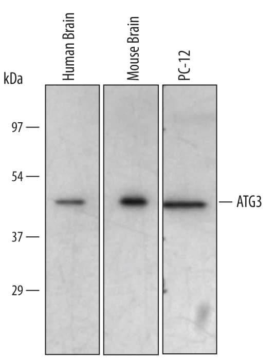
Detection of Human/Mouse/Rat ATG3 by Western Blot.
Western blot shows lysates of human brain tissue, mouse brain tissue, and PC-12 rat adrenal pheochromocytoma cell line. PVDF membrane was probed with 1 µg/mL of Sheep Anti-Human/Mouse/Rat ATG3 Antigen Affinity-purified Polyclonal Antibody (Catalog # AF5450) followed by HRP-conjugated Anti-Sheep IgG Secondary Antibody (Catalog # HAF016). A specific band was detected for ATG3 at approximately 45 kDa (as indicated). This experiment was conducted under reducing conditions and using Immunoblot Buffer Group 2.ATG3 in RAW 264.7 Mouse Cell Line.
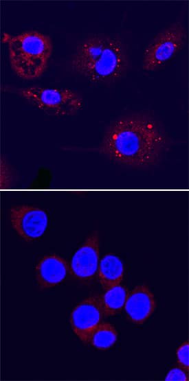
ATG3 was detected in immersion fixed RAW 264.7 mouse monocyte/macrophage cell line, untreated (lower panel) or treated with LPS (upper panel), using Sheep Anti-Human/Mouse/Rat ATG3 Antigen Affinity-purified Polyclonal Antibody (Catalog # AF5450) at 5 µg/mL for 3 hours at room temperature. Cells were stained using the NorthernLights™ 557-conjugated Anti-Sheep IgG Secondary Antibody (red; Catalog # NL010) and counterstained with DAPI (blue). Specific staining was localized to autophagosomes. View our protocol for Fluorescent ICC Staining of Non-adherent Cells.Applications for Human/Mouse/Rat ATG3 Antibody
Immunocytochemistry
Sample: Immersion fixed RAW264.7 mouse monocyte/macrophage cell line
Western Blot
Sample: Human brain tissue, mouse brain tissue, and PC-12 rat adrenal pheochromocytoma cell line
Formulation, Preparation, and Storage
Purification
Reconstitution
Formulation
Shipping
Stability & Storage
- 12 months from date of receipt, -20 to -70 °C as supplied.
- 1 month, 2 to 8 °C under sterile conditions after reconstitution.
- 6 months, -20 to -70 °C under sterile conditions after reconstitution.
Background: ATG3
ATG3 (autophagy-related protein 3; also APG3-like and PC3-96) is a ubiquitous 45 kDa member of the ATG3 family of proteins. It functions as an E2-like enzyme during the initial stages of autophagosome formation by catalyzing the transfer of ATG7-bound ATG8 (known as LC3, GATE16 and GABA-RAP in mammals) to phosphatidylethanolamine, critical for autophagy. Human ATG3 is 314 amino acids in length and contains an active site at Cys264 that forms a thiol ester bond with the C-terminal Gly of ATG8. There are multiple potential isoform variants. Three show a 22, 28 and 35 aa substitution for aa 289‑314, respectively, while a fourth shows an alternate start site at Met88 that may be accompanied by one of the afore mentioned substitutions. Over aa 1‑287, human ATG3 shares 97.9% aa identity with mouse ATG3.
Long Name
Alternate Names
Gene Symbol
UniProt
Additional ATG3 Products
Product Documents for Human/Mouse/Rat ATG3 Antibody
Product Specific Notices for Human/Mouse/Rat ATG3 Antibody
For research use only

