Human/Mouse/Rat BAK Antibody
R&D Systems, part of Bio-Techne | Catalog # AF8161

Key Product Details
Species Reactivity
Applications
Label
Antibody Source
Product Specifications
Immunogen
Pro20-Asn124
Accession # Q16611
Specificity
Clonality
Host
Isotype
Scientific Data Images for Human/Mouse/Rat BAK Antibody
Detection of Human/Mouse/Rat BAK by Western Blot.
Western blot shows lysates of HEK293 human embryonic kidney cell line, CH-1 mouse B cell lymphoma cell line, and PC-12 rat adrenal pheochromocytoma cell line. PVDF membrane was probed with 1 µg/mL of Human/Mouse/Rat BAK Antigen Affinity-purified Polyclonal Antibody (Catalog # AF8161) followed by HRP-conjugated Anti-Goat IgG Secondary Antibody (HAF109). A specific band was detected for BAK at approximately 29 kDa (as indicated). This experiment was conducted under reducing conditions and using Immunoblot Buffer Group 2.BAK in HEK293 Human Cell Line.
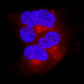
BAK was detected in immersion fixed HEK293 human embryonic kidney cell line using Goat Anti-Human/Mouse/Rat BAK Antigen Affinity-purified Polyclonal Antibody (Catalog # AF8161) at 15 µg/mL for 3 hours at room temperature. Cells were stained using the NorthernLights™ 557-conjugated Anti-Goat IgG Secondary Antibody (red; NL001) and counterstained with DAPI (blue). Specific staining was localized to cytoplasm. View our protocol for Fluorescent ICC Staining of Cells on Coverslips.Detection of BAK in Human Colon.
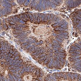
BAK was detected in immersion fixed paraffin-embedded sections of Human Colon using Goat Anti-Human/Mouse/Rat BAK Antigen Affinity-purified Polyclonal Antibody (Catalog # AF8161) at 5 µg/mL for 1 hour at room temperature followed by incubation with the Anti-Goat IgG VisUCyte™ HRP Polymer Antibody (Catalog # VC004). Before incubation with the primary antibody, tissue was subjected to heat-induced epitope retrieval using VisUCyte Antigen Retrieval Reagent-Basic (Catalog # VCTS021). Tissue was stained using DAB (brown) and counterstained with hematoxylin (blue). Specific staining was localized to cell membrane in epithelial cells in mucosal glands. View our protocol for IHC Staining with VisUCyte HRP Polymer Detection Reagents.Applications for Human/Mouse/Rat BAK Antibody
Immunocytochemistry
Sample: Immersion fixed HEK293 human embryonic kidney cell line
Immunohistochemistry
Sample: Immersion fixed paraffin-embedded sections of Human Colon
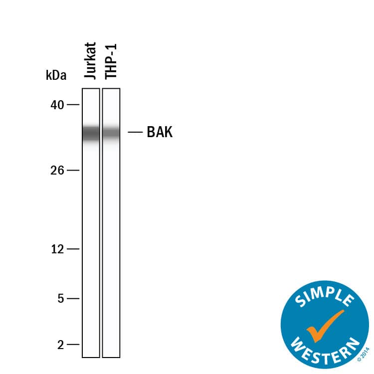
Simple Western
Sample: Jurkat human acute T cell leukemia cell line and THP-1 human acute monocytic leukemia cell line
Western Blot
Sample: HEK293 human embryonic kidney cell line, CH-1 mouse B cell lymphoma cell line, and PC-12 rat adrenal pheochromocytoma cell line
Formulation, Preparation, and Storage
Purification
Reconstitution
Formulation
*Small pack size (-SP) is supplied either lyophilized or as a 0.2 µm filtered solution in PBS.
Shipping
Stability & Storage
- 12 months from date of receipt, -20 to -70 °C as supplied.
- 1 month, 2 to 8 °C under sterile conditions after reconstitution.
- 6 months, -20 to -70 °C under sterile conditions after reconstitution.
Background: BAK
BAK (Bcl-2 homologous antagonist/killer; also BAK1) is a 25‑30 kDa member of the BCL-2 family of proteins. It is widely expressed, and participates in the apoptotic cycle. BAK is an outer mitochondrial membrane protein that is inactive as a Zn-dependent homodimer. Upon activation by p53 or tBID, BAK oligomerizes, creating a pore in the mitochondrial membrane and allowing for cytochrome C release. Human BAK is 211 amino acids (aa) in length and contains three BCL-2 homology domains (aa 74‑88, 117 ‑136 and 169‑184), a Zn-binding region (aa 160‑166) and a C-terminal transmembrane segment (aa 188‑205). Amino acids 67‑94 mediate oligomerization of BAK. There are two potential isoform variants; one shows an alternate start site at Met96, while a second shows a deletion of aa 46‑66. Over amino acids 20‑124, human BAK shares 76% aa identity with mouse BAK.
Long Name
Alternate Names
Gene Symbol
UniProt
Additional BAK Products
Product Documents for Human/Mouse/Rat BAK Antibody
Product Specific Notices for Human/Mouse/Rat BAK Antibody
For research use only


