Human/Mouse/Rat BAT3/BAG6 Antibody
R&D Systems, part of Bio-Techne | Catalog # AF6438

Key Product Details
Species Reactivity
Validated:
Cited:
Applications
Validated:
Cited:
Label
Antibody Source
Product Specifications
Immunogen
Met1-Gln190
Accession # P46379
Specificity
Clonality
Host
Isotype
Scientific Data Images for Human/Mouse/Rat BAT3/BAG6 Antibody
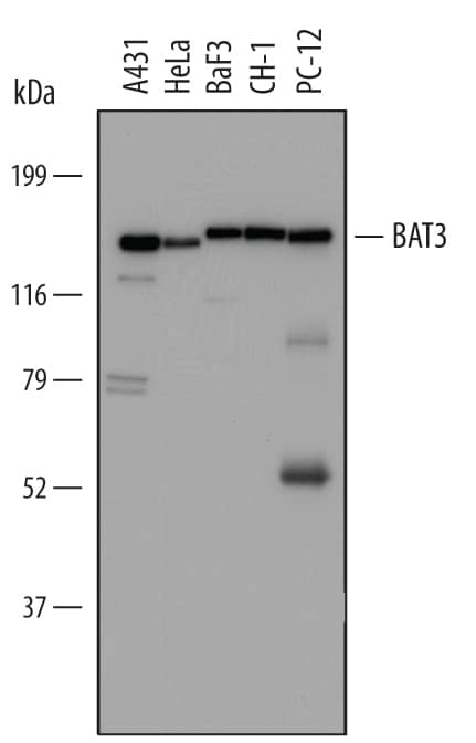
Detection of Human, Mouse, and Rat BAT3/BAG6 by Western Blot.
Western blot shows lysates of A431 human epithelial carcinoma cell line, HeLa human cervical epithelial carcinoma cell line, BaF3 mouse pro-B cell line, CH-1 mouse B cell lymphoma cell line, and PC-12 rat adrenal pheochromocytoma cell line. PVDF Membrane was probed with 1 µg/mL of Sheep Anti-Human/Mouse/Rat BAT3/BAG6 Antigen Affinity-purified Polyclonal Antibody (Catalog # AF6438) followed by HRP-conjugated Anti-Sheep IgG Secondary Antibody (Catalog # HAF016). A specific band was detected for BAT3/BAG6 at approximately 150 kDa (as indicated). This experiment was conducted under reducing conditions and using Immunoblot Buffer Group 1.BAT3/BAG6 in Human Dendritic Cells.
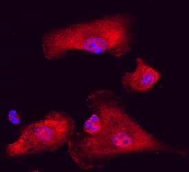
BAT3/BAG6 was detected in immersion fixed immature human dendritic cells using Sheep Anti-Human/Mouse/Rat BAT3/BAG6 Affinity-purified Polyclonal Antibody (Catalog # AF6438) at 10 µg/mL for 3 hours at room temperature. Cells were stained using the NorthernLights™ 557-conjugated Anti-Sheep IgG Secondary Antibody (red; Catalog # NL010) and counter-stained with DAPI (blue). Specific staining was localized to nuclei and cytoplasm. View our protocol for Fluorescent ICC Staining of Cells on Coverslips.Applications for Human/Mouse/Rat BAT3/BAG6 Antibody
Immunocytochemistry
Sample: Immersion fixed immature human dendritic cells
Western Blot
Sample: A431 human epithelial carcinoma cell line, HeLa human cervical epithelial carcinoma cell line, BaF3 mouse pro-B cell line, CH‑1 mouse B cell lymphoma cell line, and PC‑12 rat adrenal pheochromocytoma cell line
Formulation, Preparation, and Storage
Purification
Reconstitution
Formulation
Shipping
Stability & Storage
- 12 months from date of receipt, -20 to -70 °C as supplied.
- 1 month, 2 to 8 °C under sterile conditions after reconstitution.
- 6 months, -20 to -70 °C under sterile conditions after reconstitution.
Background: BAT3/BAG6
BAT3 (HLA-B-associated transcript 3; also BAG6, BAG family molecular chaperone regulator 6, Protein G3 and Scythe) is a multifunctional molecule that is not easily placed into known protein families. Although its predicted MW is 119 kDa, it runs anomalously in SDS-PAGE at 150-170 kDa. BAT3 is found in diverse cell types such as dendritic cells, hepatocytes, renal epithelial cells and tumor cells. It is found extracellularly where it is complexed to Hsp70, and activates the NKp30 receptor expressed on NK cells. Intracellularly, it binds to TGF-beta receptors I and II where, upon receptor ligation, it translocates to the nucleus and activates TGF-beta target genes. Human BAT3 is 1132 amino acids (aa) in length. It contains one Ub-like domain (aa 17-77), two Pro-rich regions (aa 195-273 and 394-681), an NLS (aa 1030-1052) plus a BAG domain (aa 1053-1101). BAT3 is phosphorylated on multiple Ser/Thr residues. Caspase 3 will cleave BAT3 after Asp1001, generating a 20 kDa C-terminal fragment. There are also multiple splice variants. Two of them share an absence of aa 185-190 that may be accompanied by a 36 aa insertion after Gly489. Over aa 1-190, human BAT3 shares 93% aa identity with mouse BAT3.
Long Name
Alternate Names
Gene Symbol
UniProt
Additional BAT3/BAG6 Products
Product Documents for Human/Mouse/Rat BAT3/BAG6 Antibody
Product Specific Notices for Human/Mouse/Rat BAT3/BAG6 Antibody
For research use only

