Human/Mouse/Rat beta-Actin Antibody Best Seller
R&D Systems, part of Bio-Techne | Catalog # MAB8929

Key Product Details
Validated by
Biological Validation
Species Reactivity
Validated:
Human, Mouse, Rat
Cited:
Human, Mouse, Rat, Hamster - Mesocricetus auratus (Golden Hamster), N/A, Transgenic Mouse
Applications
Validated:
Immunocytochemistry, Simple Western, Western Blot
Cited:
Immunocytochemistry, Immunoprecipitation, Simple Western, Western Blot
Label
Unconjugated
Antibody Source
Monoclonal Mouse IgG1 Clone # 937215
Product Specifications
Immunogen
Peptide containing a sequence at the N-terminus of human beta‑Actin
Accession # P60709
Accession # P60709
Specificity
Detects human, mouse, and rat beta‑Actin in Western blots.
Clonality
Monoclonal
Host
Mouse
Isotype
IgG1
Scientific Data Images for Human/Mouse/Rat beta-Actin Antibody
Detection of Human, Mouse, and Rat beta‑Actin by Western Blot.
Western blot shows lysates of A431 human epithelial carcinoma cell line, C2C12 mouse myoblast cell line, and Rat-2 rat embryonic fibroblast cell line. PVDF membrane was probed with 0.01 µg/mL of Mouse Anti-Human/Mouse/Rat beta-Actin Monoclonal Antibody (Catalog # MAB8929) followed by HRP-conjugated Anti-Mouse IgG Secondary Antibody (Catalog # HAF018). A specific band was detected for beta-Actin at approximately 45 kDa (as indicated). This experiment was conducted under reducing conditions and using Immunoblot Buffer Group 1.beta‑Actin in NIH‑3T3 Mouse Cell Line.
beta-Actin was detected in immersion fixed NIH-3T3 mouse embryonic fibroblast cell line using Mouse Anti-Human/Mouse/Rat beta-Actin Monoclonal Antibody (Catalog # MAB8929) at 0.1 µg/mL for 3 hours at room temperature. Cells were stained using the NorthernLights™ 557-conjugated Anti-Mouse IgG Secondary Antibody (red; Catalog # NL007) and counterstained with DAPI (blue). Cells were fixated in methanol. Specific staining was localized to cytoskeleton.Detection of Human beta‑Actin by Simple WesternTM.
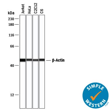
Simple Western lane view shows lysate of A431 human epithelial carcinoma cell line, loaded at 0.2 mg/mL. A specific band was detected for beta‑Actin at approximately 49 kDa (as indicated) using 1 µg/mL of Mouse Anti-Human/Mouse/Rat beta‑Actin Monoclonal Antibody (Catalog # MAB8929). This experiment was conducted under reducing conditions and using the 12-230 kDa separation system.Applications for Human/Mouse/Rat beta-Actin Antibody
Application
Recommended Usage
Immunocytochemistry
0.1-25 µg/mL
Sample: Immersion fixed NIH-3T3 mouse embryonic fibroblast cell line
Sample: Immersion fixed NIH-3T3 mouse embryonic fibroblast cell line
Simple Western
1 µg/mL
Sample: A431 human epithelial carcinoma cell line, Jurkat human acute T cell leukemia cell line, HeLa human cervical epithelial carcinoma cell line, C2C12 mouse myoblast cell line, and C6 rat glioma cell line
Sample: A431 human epithelial carcinoma cell line, Jurkat human acute T cell leukemia cell line, HeLa human cervical epithelial carcinoma cell line, C2C12 mouse myoblast cell line, and C6 rat glioma cell line
Western Blot
0.01 µg/mL
Sample: A431 human epithelial carcinoma cell line, C2C12 mouse myoblast cell line, and Rat‑2 rat embryonic fibroblast cell line
Sample: A431 human epithelial carcinoma cell line, C2C12 mouse myoblast cell line, and Rat‑2 rat embryonic fibroblast cell line
Reviewed Applications
Read 14 reviews rated 4.8 using MAB8929 in the following applications:
Formulation, Preparation, and Storage
Purification
Protein A or G purified from hybridoma culture supernatant
Reconstitution
Reconstitute at 0.5 mg/mL in sterile PBS. For liquid material, refer to CoA for concentration.
Formulation
Lyophilized from a 0.2 μm filtered solution in PBS with Trehalose. *Small pack size (SP) is supplied either lyophilized or as a 0.2 µm filtered solution in PBS.
Shipping
Lyophilized product is shipped at ambient temperature. Liquid small pack size (-SP) is shipped with polar packs. Upon receipt, store immediately at the temperature recommended below.
Stability & Storage
Use a manual defrost freezer and avoid repeated freeze-thaw cycles.
- 12 months from date of receipt, -20 to -70 °C as supplied.
- 1 month, 2 to 8 °C under sterile conditions after reconstitution.
- 6 months, -20 to -70 °C under sterile conditions after reconstitution.
Background: beta-Actin
( gamma-Smooth Muscle, gamma-non-muscle) isoforms. Non-muscle beta- and gamma-actin are also known as cytoplasmic actin.
Alternate Names
ACTB, betaActin
Gene Symbol
ACTB
UniProt
Additional beta-Actin Products
Product Documents for Human/Mouse/Rat beta-Actin Antibody
Product Specific Notices for Human/Mouse/Rat beta-Actin Antibody
For research use only
Loading...
Loading...
Loading...
Loading...
Loading...


