Human/Mouse/Rat Coactosin-like Protein 1/COTL1 Antibody
R&D Systems, part of Bio-Techne | Catalog # AF7865

Key Product Details
Species Reactivity
Applications
Label
Antibody Source
Product Specifications
Immunogen
Ala2-Glu142
Accession # Q14019
Specificity
Clonality
Host
Isotype
Scientific Data Images for Human/Mouse/Rat Coactosin-like Protein 1/COTL1 Antibody
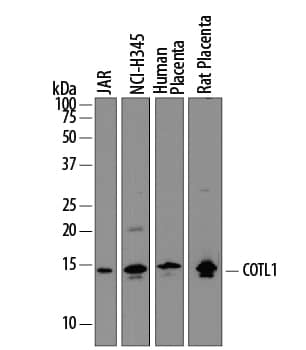
Detection of Human and Rat Coactosin-like Protein 1/COTL1 by Western Blot.
Western blot shows lysates of JAR human choriocarcinoma cell line, NCI-H345 human small cell lung carcinoma cell line, human placenta tissue, and rat placenta tissue. PVDF membrane was probed with 1 µg/mL of Sheep Anti-Human/Mouse/Rat Coactosin-like Protein 1/COTL1 Antigen Affinity-purified Polyclonal Antibody (Catalog # AF7865) followed by HRP-conjugated Anti-Sheep IgG Secondary Antibody (Catalog # HAF016). A specific band was detected for Coactosin-like Protein 1/COTL1 at approximately 15 kDa (as indicated). This experiment was conducted under reducing conditions and using Immunoblot Buffer Group 1.Coactosin-like Protein 1/COTL1 in NCI‑H128 Human Cell Line.
Coactosin-like Protein 1/COTL1 was detected in immersion fixed NCI-H128 human small cell lung carcinoma cell line using Sheep Anti-Human/Mouse/Rat Coactosin-like Protein 1/COTL1 Antigen Affinity-purified Polyclonal Antibody (Catalog # AF7865) at 10 µg/mL for 3 hours at room temperature. Cells were stained using the NorthernLights™ 557-conjugated Anti-Sheep IgG Secondary Antibody (red; Catalog # NL010) and counterstained with DAPI (blue). Specific staining was localized to cytoplasm. View our protocol for Fluorescent ICC Staining of Non-adherent Cells.Applications for Human/Mouse/Rat Coactosin-like Protein 1/COTL1 Antibody
Immunocytochemistry
Sample: Immersion fixed NCI‑H128 human small cell lung carcinoma cell line
Western Blot
Sample: JAR human choriocarcinoma cell line, NCI‑H345 human small cell lung carcinoma cell line, human placenta tissue, and rat placenta tissue
Formulation, Preparation, and Storage
Purification
Reconstitution
Formulation
Shipping
Stability & Storage
- 12 months from date of receipt, -20 to -70 °C as supplied.
- 1 month, 2 to 8 °C under sterile conditions after reconstitution.
- 6 months, -20 to -70 °C under sterile conditions after reconstitution.
Background: Coactosin-like Protein 1/CotL1
COTL1 (Coactosin-like Protein) is both a cytoplasmic and plasma-appearing 15-16 kDa member of the coactosin subfamily, ADF/Actin Depolymerizing Factor family of actin-binding proteins. It is widely expressed, and found in cell types such as neutrophils, and tissues such as placenta, lung and kidney. Functionally, COTL1 interacts noncovalently with both F-actin and 5-lipoxygenase/5LO. These interactions appear to be mutually exclusive. A COTL1:F-actin interaction leads to actin binding without actin polymerization, while a 5LO:COTL1 interaction has two potential outcomes; first, 5LO sequesters COTL1, leading to a failure of actin binding, and second, COTL1 can serve as a scaffold for 5LO activity, facilitating the production of either 5HPETE or LTA4. Human COTL1 is 142 amino acids (aa) in length. It is principally composed of one ADF-H domain (aa 2-130) that possesses a utilized phosphorylation site at Ser115, and two acetylation sites at Lys102 and Lys126. COTL1 may form noncovalent homodimers and oligomers, but not when complexed to F-actin. There is one potential isoform variant that shows a 106 aa substitution for aa 1-53. Full-length human and mouse COTL1 share 95% aa sequence identity.
Alternate Names
Gene Symbol
UniProt
Additional Coactosin-like Protein 1/CotL1 Products
Product Documents for Human/Mouse/Rat Coactosin-like Protein 1/COTL1 Antibody
Product Specific Notices for Human/Mouse/Rat Coactosin-like Protein 1/COTL1 Antibody
For research use only

