Human/Mouse/Rat DOK7 Antibody
R&D Systems, part of Bio-Techne | Catalog # AF6398

Key Product Details
Validated by
Species Reactivity
Validated:
Cited:
Applications
Validated:
Cited:
Label
Antibody Source
Product Specifications
Immunogen
Ala179-Pro299
Accession # Q18PE1
Specificity
Clonality
Host
Isotype
Scientific Data Images for Human/Mouse/Rat DOK7 Antibody
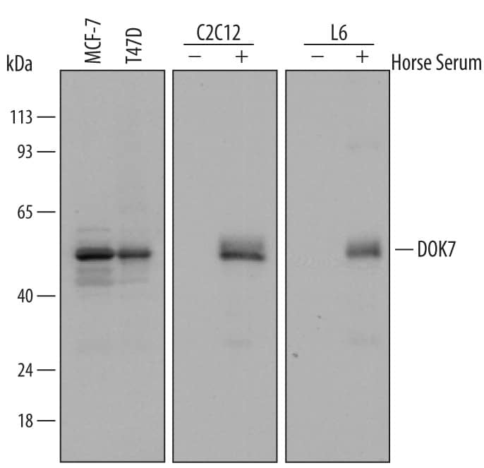
Detection of Human, Mouse, and Rat DOK7 by Western Blot.
Western blot shows lysates of untreated MCF-7 human breast cancer cell line and T47D human breast cancer cell line and C2C12 mouse myoblast cell line and L6 rat myoblast cell line untreated (-) or treated (+) with 2% horse serum for 6 days. PVDF Membrane was probed with 0.5 µg/mL of Goat Anti-Human/Mouse/Rat DOK7 Antigen Affinity-purified Polyclonal Antibody (Catalog # AF6398) followed by HRP-conjugated Anti-Goat IgG Secondary Antibody (Catalog # HAF109). A specific band was detected for DOK7 at approximately 55 kDa (as indicated). This experiment was conducted under reducing conditions and using Immunoblot Buffer Group 2.Detection of Human DOK7 by Simple WesternTM.
Simple Western lane view shows lysates of MCF-7 human breast cancer cell line and T47D human breast cancer cell line, loaded at 0.2 mg/mL. A specific band was detected for DOK7 at approximately 60 & 62 kDa (as indicated) using 5 µg/mL of Goat Anti-Human/Mouse/Rat DOK7 Antigen Affinity-purified Polyclonal Antibody (Catalog # AF6398) followed by 1:50 dilution of HRP-conjugated Anti-Goat IgG Secondary Antibody (Catalog # HAF109). This experiment was conducted under reducing conditions and using the 12-230 kDa separation system.Detection of Mouse DOK7 by Western Blot
Sorbs1 is enriched at AChR aggregates, and Sorbs1 RNAi blocks AChR clustering in vitro. (A) Treatment of myotubes with siRNA directed against Sorbs1 blocks AChR clustering in C2C12 cells. Montages containing sixteen fields at a magnification of 10× were analyzed with ImageJ software (NIH). (B) Sorbs1 siRNA significantly reduces Sorbs1 protein expression in myotubes. (C) Sorbs1 protein is highly enriched at sites where AChRs aggregate. (D) Agrin stimulates tyrosine phosphorylation of MuSK and Dok-7 at similar levels in myotubes treated with Sorbs1 siRNA. Image collected and cropped by CiteAb from the following publication (https://pubmed.ncbi.nlm.nih.gov/26527617), licensed under a CC-BY license. Not internally tested by R&D Systems.Applications for Human/Mouse/Rat DOK7 Antibody
Simple Western
Sample: MCF‑7 human breast cancer cell line and T47D human breast cancer cell line
Western Blot
Sample: MCF‑7 human breast cancer cell line and T47D human breast cancer cell line (untreated) and C2C12 mouse myoblast cell line and L6 rat myoblast cell line treated with horse serum
Formulation, Preparation, and Storage
Purification
Reconstitution
Formulation
Shipping
Stability & Storage
- 12 months from date of receipt, -20 to -70 °C as supplied.
- 1 month, 2 to 8 °C under sterile conditions after reconstitution.
- 6 months, -20 to -70 °C under sterile conditions after reconstitution.
Background: DOK7
DOK7 (Downstream of kinase 7) is a 55 kDa member of the DOK family of cytoplasmic adaptor proteins. It links the acetylcholine receptor and the receptor tyrosine kinase MuSK in skeletal and cardiac muscle. Mutations can cause familial myasthenic syndromes. The 504 amino acid (aa) human DOK7 contains pleckstrin homology (aa 7‑109) and phosphotyrosine-binding (PTK, aa 125‑236) and SH2 domains and a C‑terminal nuclear export signal. Splicing isoforms of 255, 608, and 366 aa diverge at aa 175 or 500, or have 40 divergent aa replacing aa 1‑178, respectively. Within aa 179‑299, human DOK7 shares 92% and 93% aa identity with mouse and rat DOK7, respectively.
Long Name
Alternate Names
Gene Symbol
UniProt
Additional DOK7 Products
Product Documents for Human/Mouse/Rat DOK7 Antibody
Product Specific Notices for Human/Mouse/Rat DOK7 Antibody
For research use only


