Human/Mouse/Rat EMP/MAEA Antibody
R&D Systems, part of Bio-Techne | Catalog # AF7288

Key Product Details
Species Reactivity
Validated:
Human, Mouse, Rat
Cited:
Human, Mouse
Applications
Validated:
Immunocytochemistry, Western Blot
Cited:
Immunoprecipitation, Western Blot
Label
Unconjugated
Antibody Source
Polyclonal Sheep IgG
Product Specifications
Immunogen
E. coli-derived recombinant human EMP/MAEA
Met12-Ser66
Accession # Q7L5Y9
Met12-Ser66
Accession # Q7L5Y9
Specificity
Detects human, mouse and rat EMP/MAEA in Western blots. Detects recombinant human EMP/MAEA in direct ELISAs.
Clonality
Polyclonal
Host
Sheep
Isotype
IgG
Scientific Data Images for Human/Mouse/Rat EMP/MAEA Antibody
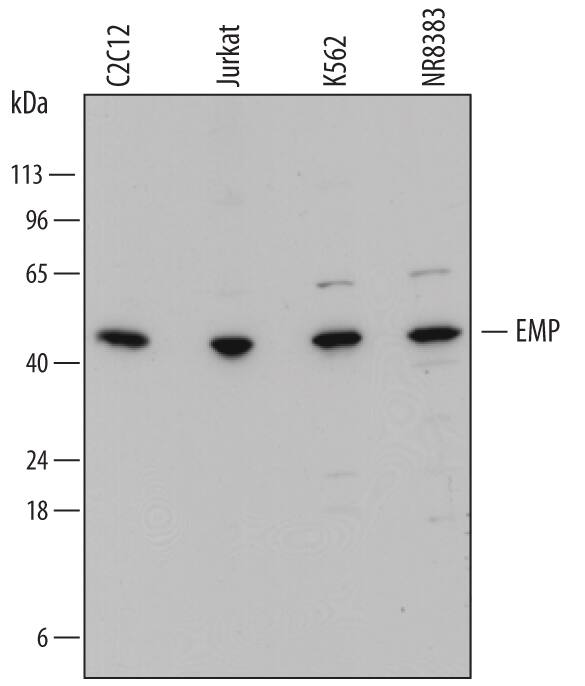
Detection of Human, Mouse, and Rat EMP/MAEA by Western Blot.
Western blot shows lysates of C2C12 mouse myoblast cell line, Jurkat human acute T cell leukemia cell line, K562 human chronic myelogenous leukemia cell line, and NR8383 rat alveolar macrophage cell line. PVDF membrane was probed with 1 µg/mL of Sheep Anti-Human EMP/MAEA Antigen Affinity-purified Polyclonal Antibody (Catalog # AF7288) followed by HRP-conjugated Anti-Sheep IgG Secondary Antibody (Catalog # HAF016). A specific band was detected for EMP/MAEA at approximately 45 kDa (as indicated). This experiment was conducted under reducing conditions and using Immunoblot Buffer Group 1.EMP/MAEA in Jurkat Human Cell Line.
EMP/MAEA was detected in immersion fixed Jurkat human acute T cell leukemia cell line using Sheep Anti-Human EMP/MAEA Antigen Affinity-purified Polyclonal Antibody (Catalog # AF7288) at 10 µg/mL for 3 hours at room temperature. Cells were stained using the NorthernLights™ 557-conjugated Anti-Sheep IgG Secondary Antibody (red; Catalog # NL010) and counterstained with DAPI (blue). Specific staining was localized to cytoplasm and nuclei. View our protocol for Fluorescent ICC Staining of Non-adherent Cells.Detection of Mouse EMP/MAEA by Western Blot
V5-HA-tagged RanBP9 maintains its ability to interact with known members of the CTLH complex and Nucleolin. RanBP9 WT and TT immortalized Mouse Embryonic Fibroblasts (MEFs) were cultured in standard conditions and protein lysates were obtained. Resin conjugated with alphaHA antibodies was used to immunoprecipitate RanBP9-TT protein. IPed fractions and 5% of input were run on gels to generate 5 different membranes that were probed with the indicated antibodies by WB. Vinculin is used as loading control. Shown results are representative of two independent experiments (biological replicates). Image collected and cropped by CiteAb from the following publication (https://pubmed.ncbi.nlm.nih.gov/32346083), licensed under a CC-BY license. Not internally tested by R&D Systems.Applications for Human/Mouse/Rat EMP/MAEA Antibody
Application
Recommended Usage
Immunocytochemistry
5-15 µg/mL
Sample: Immersion fixed Jurkat human acute T cell leukemia cell line
Sample: Immersion fixed Jurkat human acute T cell leukemia cell line
Western Blot
1 µg/mL
Sample: C2C12 mouse myoblast cell line, Jurkat human acute T cell leukemia cell line, K562 human chronic myelogenous leukemia cell line, and NR8383 rat alveolar macrophage cell line
Sample: C2C12 mouse myoblast cell line, Jurkat human acute T cell leukemia cell line, K562 human chronic myelogenous leukemia cell line, and NR8383 rat alveolar macrophage cell line
Formulation, Preparation, and Storage
Purification
Antigen Affinity-purified
Reconstitution
Sterile PBS to a final concentration of 0.2 mg/mL. For liquid material, refer to CoA for concentration.
Formulation
Lyophilized from a 0.2 μm filtered solution in PBS with Trehalose. *Small pack size (SP) is supplied either lyophilized or as a 0.2 µm filtered solution in PBS.
Shipping
Lyophilized product is shipped at ambient temperature. Liquid small pack size (-SP) is shipped with polar packs. Upon receipt, store immediately at the temperature recommended below.
Stability & Storage
Use a manual defrost freezer and avoid repeated freeze-thaw cycles.
- 12 months from date of receipt, -20 to -70 °C as supplied.
- 1 month, 2 to 8 °C under sterile conditions after reconstitution.
- 6 months, -20 to -70 °C under sterile conditions after reconstitution.
Background: EMP/MAEA
Long Name
Erythroblast Macrophage Protein/Macrophage Erythroblast Attacher
Alternate Names
EMLP, HLC-10, MAEA, PIG5
Gene Symbol
MAEA
UniProt
Additional EMP/MAEA Products
Product Documents for Human/Mouse/Rat EMP/MAEA Antibody
Product Specific Notices for Human/Mouse/Rat EMP/MAEA Antibody
For research use only
Loading...
Loading...
Loading...
Loading...


