Human/Mouse/Rat RAGE/AGER Antibody
R&D Systems, part of Bio-Techne | Catalog # AF1145

Key Product Details
Validated by
Species Reactivity
Validated:
Cited:
Applications
Validated:
Cited:
Label
Antibody Source
Product Specifications
Immunogen
Gln24-Ala344
Accession # Q15109
Specificity
Clonality
Host
Isotype
Endotoxin Level
Scientific Data Images for Human/Mouse/Rat RAGE/AGER Antibody
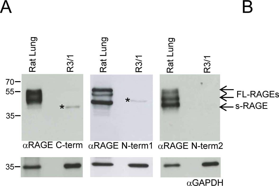
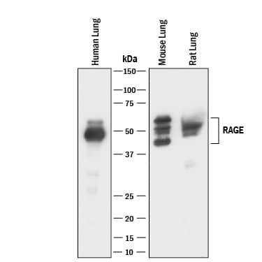
Detection of Human, Mouse, and Rat RAGE by Western Blot.
Western blot shows lysates of human, mouse, and rat lung tissue. PVDF membrane was probed with 1 µg/mL of Goat Anti-Human/Mouse/Rat RAGE Antigen Affinity-purified Polyclonal Antibody (Catalog # AF1145) followed by HRP-conjugated Anti-Goat IgG Secondary Antibody (Catalog # HAF017). Specific bands were detected for RAGE at approximately 45-55 kDa (as indicated). This experiment was conducted under reducing conditions and using Immunoblot Buffer Group 1.Detection of Human RAGE by Western Blot.
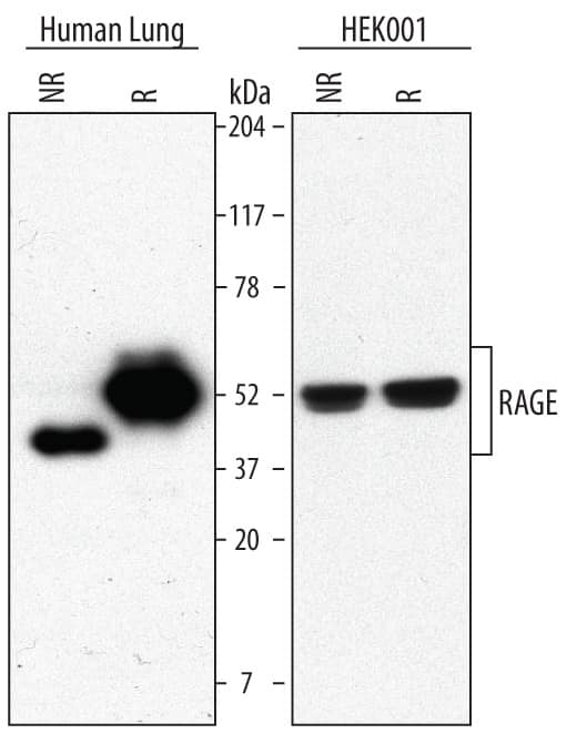
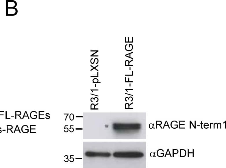
Western blot shows lysates of human lung tissue and HEK001 human epidermal keratinocyte cell line. PVDF Membrane was probed with 1 µg/mL of Goat Anti-Human/Mouse/Rat RAGE Antigen Affinity-purified Polyclonal Antibody (Catalog # AF1145) followed by HRP-conjugated Anti-Goat IgG Secondary Antibody (Catalog # HAF017). Specific bands were detected for RAGE at approximately 43 kDa under non-reducing (NR) conditions and 50 kDa under reducing (R) conditions (as indicated). This experiment was conducted using Immunoblot Buffer Group 5.RAGE in Human Alzheimer's Disease Brain.
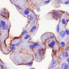
RAGE was detected in immersion fixed paraffin-embedded sections of human Alzheimer's disease brain (cerebellum) using 15 µg/mL Goat Anti-Human/Mouse/Rat RAGE Antigen Affinity-purified Polyclonal Antibody (Catalog # AF1145) overnight at 4 °C. Tissue was stained with the Anti-Goat HRP-DAB Cell & Tissue Staining Kit (brown; Catalog # CTS008) and counterstained with hematoxylin (blue). View our protocol for Chromogenic IHC Staining of Paraffin-embedded Tissue Sections.Applications for Human/Mouse/Rat RAGE/AGER Antibody
Blockade of Receptor-ligand Interaction
Immunohistochemistry
Sample: Immersion fixed paraffin-embedded sections of human Alzheimer's disease brain (cerebellum) and immersion fixed paraffin-embedded sections of human lung
Western Blot
Sample: Human, mouse, and rat lung tissue and HEK001 human epidermal keratinocyte cell line
Reviewed Applications
Read 3 reviews rated 5 using AF1145 in the following applications:
Formulation, Preparation, and Storage
Purification
Reconstitution
Formulation
Shipping
Stability & Storage
- 12 months from date of receipt, -20 to -70 °C as supplied.
- 1 month, 2 to 8 °C under sterile conditions after reconstitution.
- 6 months, -20 to -70 °C under sterile conditions after reconstitution.
Background: RAGE/AGER
Advanced glycation endproducts (AGE) are adducts formed by the non-enzymatic glycation or oxidation of macromolecules. AGE forms during aging and its formation is accelerated under pathophysiologic states such as diabetes, Alzheimer’s disease, renal failure and immune/inflammatory disorders. Receptor for Advanced Glycation Endoproducts (RAGE), named for its ability to bind AGE, is a multi-ligand receptor belonging the immunoglobulin (Ig) superfamily. Besides AGE, RAGE binds amyloid beta-peptide, S100/calgranulin family proteins, high mobility group B1 (HMGB1, also know as amphoterin) and leukocyte integrins.
The human RAGE gene encodes a 404 amino acid residues (aa) type I transmembrane glycoprotein with a 22 aa signal peptide, a 320 aa extracellular domain containing an Ig-like V-type domain and two Ig-like Ce-type domains, a 21 aa transmembrane domain and a 41 aa cytoplasmic domain. The V-type domain and the cytoplasmic domain are important for ligand binding and for intracellular signaling, respectively. Two alternative splice variants, lacking the V-type domain or the cytoplasmic tail, are known. RAGE is highly expressed in the embryonic central nervous system. In adult tissues, RAGE is expressed at low levels in multiple tissues including endothelial and smooth muscle cells, mononuclear phagocytes, pericytes, microglia, neurons, cardiac myocytes, and hepatocytes. The expression of RAGE is upregulated upon ligand interaction. Depending on the cellular context and interacting ligand, RAGE activation can trigger differential signaling pathways that affect divergent pathways of gene expression. RAGE activation modulates varied essential cellular responses (including inflammation, immunity, proliferation, cellular adhesion, and migration) that contribute to cellular dysfunction associated with chronic diseases such as diabetes, cancer, amyloidoses, and immune or inflammatory disorders.
Long Name
Alternate Names
Gene Symbol
UniProt
Additional RAGE/AGER Products
Product Documents for Human/Mouse/Rat RAGE/AGER Antibody
Product Specific Notices for Human/Mouse/Rat RAGE/AGER Antibody
For research use only
