Human/Mouse/Rat Teneurin-2 Antibody
R&D Systems, part of Bio-Techne | Catalog # AF4578

Key Product Details
Species Reactivity
Validated:
Cited:
Applications
Validated:
Cited:
Label
Antibody Source
Product Specifications
Immunogen
Met1-Lys183
Accession # AAI72353
Specificity
Clonality
Host
Isotype
Scientific Data Images for Human/Mouse/Rat Teneurin-2 Antibody
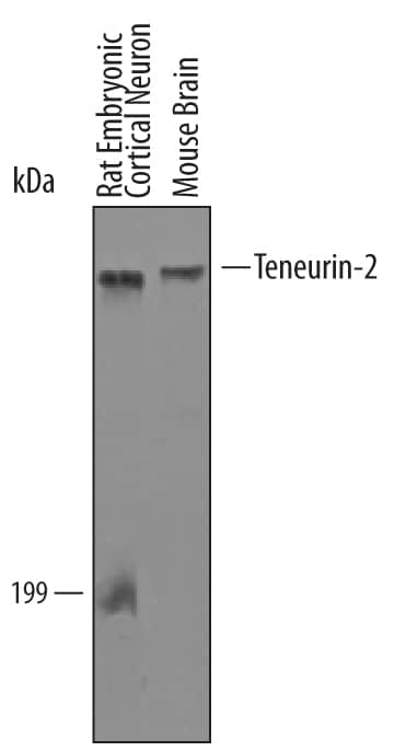
Detection of Mouse and Rat Teneurin-2 by Western blot.
Western blot shows lysates of mouse brain tissue and rat embryonic cortical neuron cells. PVDF Membrane was probed with 1 µg/mL of Human Teneurin-2 Antigen Affinity-purified Polyclonal Antibody (Catalog # AF4578) followed by HRP-conjugated Anti-Sheep IgG Secondary Antibody (Catalog # HAF016). A specific band was detected for Teneurin-2 at approximately 300 kDa (as indicated). This experiment was conducted under reducing conditions and using Immunoblot Buffer Group 8.Teneurin‑2 in Mouse Brain.
Teneurin-2 was detected in immersion fixed frozen sections of embryonic mouse brain using Sheep Anti-Human/Mouse/Rat Teneurin-2 Antigen Affinity-purified Polyclonal Antibody (Catalog # AF4578) at 10 µg/mL overnight at 4 °C. Tissue was stained using the Anti-Sheep HRP-DAB Cell & Tissue Staining Kit (brown; Catalog # CTS019) and counterstained with hematoxylin (blue). Specific staining was localized to neurons in dorsal root ganglia and cells in cartilage primordium. View our protocol for Chromogenic IHC Staining of Frozen Tissue Sections.Detection of Human/Mouse/Rat Teneurin-2 by Western Blot.
Western blot shows lysates of U251-MG human malignant glioblastoma cell line. PVDF membrane was probed with 1 µg/mL of Sheep Anti-Human/Mouse/Rat Teneurin-2 Antigen Affinity-purified Polyclonal Antibody (Catalog # AF4578) followed by HRP-conjugated Anti-Sheep IgG Secondary Antibody (Catalog # HAF016). A specific band was detected for Teneurin-2 at approximately 300 kDa (as indicated). This experiment was conducted under reducing conditions and using Western Blot Buffer Group 1.Applications for Human/Mouse/Rat Teneurin-2 Antibody
Immunohistochemistry
Sample: Immersion fixed frozen sections of embryonic mouse brain
Western Blot
Sample: Mouse brain tissue, rat embryonic cortical neuron cells and U251-MG human malignant glioblastoma cell line.
Formulation, Preparation, and Storage
Purification
Reconstitution
Formulation
Shipping
Stability & Storage
- 12 months from date of receipt, -20 to -70 °C as supplied.
- 1 month, 2 to 8 °C under sterile conditions after reconstitution.
- 6 months, -20 to -70 °C under sterile conditions after reconstitution.
Background: Teneurin-2
Teneurin-2 (also Ten-2/Ten-m2, tenascin-M2, and Ten-m/Odz2) is a 250-330 kDa member of the tenascin family, teneurin subfamily of transmembrane (TM) molecules. It is a covalently-linked homodimer that is expressed in both embryonic and adult neurons, among which are cerebellar Purkinje cells, pyramidal neurons of the hippocampus, and neurons of layers II and VI of the cerebral cortex. Teneurin-2 appears to promote neurite outgrowth and mediate cell-to-cell adhesion via homophilic interactions. Human teneurin-2 is a 2774 amino acid (aa) type II TM glycoprotein. It contains a 379 aa cytoplasmic region (aa 1-379) that contains a polySer segment (aa 331-334), plus a 2374 aa extracellular domain (ECD). The ECD possesses eight sequential EGF-like domains (aa 575-841), five NHL repeats, each of which forms a beta-propeller (aa 1272-1573), and 23 YD/TyrAsp-containing repeats that bind carbohydrates. There is a furin cleavage site after Arg528. There are three potential splice variants. One shows a deletion of aa 799-807, while two others show 46 and five aa substitutions for aa 1-167, respectively. Over aa 1-183, human teneurin-2 shares 98% aa identity with mouse teneurin-2.
Alternate Names
Gene Symbol
UniProt
Additional Teneurin-2 Products
Product Documents for Human/Mouse/Rat Teneurin-2 Antibody
Product Specific Notices for Human/Mouse/Rat Teneurin-2 Antibody
For research use only


