Human/Mouse/Rat TrkB Antibody
R&D Systems, part of Bio-Techne | Catalog # AF1494


Key Product Details
Species Reactivity
Validated:
Cited:
Applications
Validated:
Cited:
Label
Antibody Source
Product Specifications
Immunogen
Cys32-His429
Accession # P15209
Specificity
Clonality
Host
Isotype
Endotoxin Level
Scientific Data Images for Human/Mouse/Rat TrkB Antibody
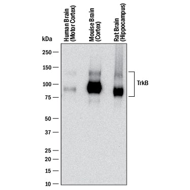
Detection of Human, Mouse, and Rat TrkB by Western Blot.
Western blot shows lysates of human brain (motor cortex) tissue, mouse brain (cortex) tissue, and rat brain (hippocampus) tissue. PVDF membrane was probed with 0.1 µg/mL of Goat Anti-Human/Mouse/Rat TrkB Antigen Affinity-purified Polyclonal Antibody (Catalog # AF1494) followed by HRP-conjugated Anti-Goat IgG Secondary Antibody (HAF017). Specific bands were detected for TrkB at approximately 95 kDa and 145 kDa (as indicated). This experiment was conducted under reducing conditions and using Immunoblot Buffer Group 1.Detection of Mouse TrkB by Western Blot.
Western blot shows lysates of mouse brain (cerebellum) tissue and mouse brain (cortex) tissue . PVDF membrane was probed with 0.5 µg/mL of Goat Anti-Human/Mouse/Rat TrkB Antigen Affinity-purified Polyclonal Antibody (Catalog # AF1494) followed by HRP-conjugated Anti-Goat IgG Secondary Antibody (HAF017). Specific bands were detected for TrkB at approximately 90-100 and 140 kDa (as indicated). This experiment was conducted under reducing conditions and using Immunoblot Buffer Group 1.TrkB in Mouse Spinal Cord.
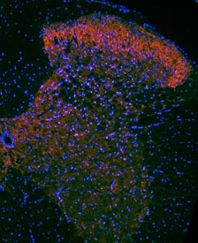
TrkB was detected in perfusion fixed frozen sections of mouse spinal cord using Goat Anti-Human/Mouse/Rat TrkB Antigen Affinity-purified Polyclonal Antibody (Catalog # AF1494) at 1.7 µg/mL overnight at 4 °C. Tissue was stained using the NorthernLights™ 557-conjugated Anti-Goat IgG Secondary Antibody (red; NL001) and counterstained with DAPI (blue). Specific staining was localized to neuronal processes and cell bodies. View our protocol for Fluorescent IHC Staining of Frozen Tissue Sections.Applications for Human/Mouse/Rat TrkB Antibody
Blockade of Receptor-ligand Interaction
Immunohistochemistry
Sample: Perfusion fixed frozen sections of mouse brain and spinal cord
Simple Western
Sample: Mouse brain (cortex) tissue
Western Blot
Sample: Human brain (motor cortex) tissue, Mouse brain (cortex) tissue, Rat brain (hippocampus) tissue, and Mouse brain (cerebellum) tissue
Reviewed Applications
Read 17 reviews rated 4.6 using AF1494 in the following applications:
Formulation, Preparation, and Storage
Purification
Reconstitution
Formulation
Shipping
Stability & Storage
- 12 months from date of receipt, -20 to -70 °C as supplied.
- 1 month, 2 to 8 °C under sterile conditions after reconstitution.
- 6 months, -20 to -70 °C under sterile conditions after reconstitution.
Background: TrkB
The neurotrophins, including NGF, BDNF, NT-3, and NT-4/5, constitute a group of structurally related, secreted proteins that play an important role in the development and function of the nervous system. The biological activities of the neurotrophins are mediated by binding to and activating two unrelated receptor types: the p75 neurotrophin receptor (p75NTR) and the Trk family of receptor tyrosine kinases (1, 2). p75NTR is a member of the tumor necrosis factor receptor superfamily (TNFRSF) and has been designated TNFRSF16. It binds all neurotrophins with low affinity to transduce cellular signaling pathways that synergize with or antagonize those activated by the Trk receptors. Three Trk family proteins, TrkA, TrkB, and TrkC, exhibiting different ligand specificities, have been identified. TrkA binds NGF and NT-3, TrkB binds BDNF, NT-3, and NT-4/5, and TrkC only binds NT-3 (1-2). All Trk family proteins share a conserved, complex subdomain organization consisting of a signal peptide, two cysteine-rich domains, a cluster of three leucine-rich motifs, and two immunoglobulin-like domains in the extracellular region, as well as an intracellular region that contains the tyrosine kinase domain (3). Natural splice variants of the different Trks, lacking the first cysteine-rich domain, the first and second or all three of the leucine-rich motifs, or the tyrosine kinase domain, have been described (4). At the protein sequence level, human and mouse TrkB show 94% amino acid sequence identity (5-6). The proteins also exhibit cross-species activity. The primary location of TrkB expression is in the central and peripheral nervous systems. Low level TrkB expression has also been observed in a wide variety of tissues outside the nervous system (6).
References
- Huang, E.J. and L.F. Reichardt (2003) Annu. Rev. Biochem. 72:609.
- Dechant, G. (2001) Cell Tissue Res. 305:229.
- Schneider, R. and M. Schweiger (1991) Oncogene 6:1807.
- Ninkina, N. et al. (1997) J. Biol. Chem. 272:13019.
- Nakagawara A. et al. (1995) Genomics. 25:538.
- Klein, R. et al. (1989) EMBO J. 8:3701.
Long Name
Alternate Names
Gene Symbol
UniProt
Additional TrkB Products
Product Documents for Human/Mouse/Rat TrkB Antibody
Product Specific Notices for Human/Mouse/Rat TrkB Antibody
For research use only


