Human/Mouse S100A6 Antibody
R&D Systems, part of Bio-Techne | Catalog # AF4584


Key Product Details
Validated by
Species Reactivity
Validated:
Cited:
Applications
Validated:
Cited:
Label
Antibody Source
Product Specifications
Immunogen
Met1-Lys89
Accession # P14069
Specificity
Clonality
Host
Isotype
Scientific Data Images for Human/Mouse S100A6 Antibody
Detection of Mouse S100A6 by Western Blot.
Western blot shows lysates of RAW 264.7 mouse monocyte/macrophage cell line and NIH-3T3 mouse embryonic fibroblast cell line. PVDF membrane was probed with 1 µg/mL of Sheep Anti-Human/ Mouse S100A6 Antigen Affinity-purified Polyclonal Antibody (Catalog # AF4584) followed by HRP-conjugated Anti-Sheep IgG Secondary Antibody (Catalog # HAF016). A specific band was detected for S100A6 at approximately 10 kDa (as indicated). This experiment was conducted under reducing conditions and using Immunoblot Buffer Group 8.S100A6 in Human Kidney.
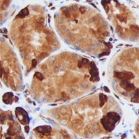
S100A6 was detected in immersion fixed paraffin-embedded sections of human kidney using Sheep Anti-Human/Mouse S100A6 Antigen Affinity-purified Polyclonal Antibody (Catalog # AF4584) at 3 µg/mL overnight at 4 °C. Before incubation with the primary antibody, tissue was subjected to heat-induced epitope retrieval using Antigen Retrieval Reagent-Basic (Catalog # CTS013). Tissue was stained using the Anti-Sheep HRP-DAB Cell & Tissue Staining Kit (brown; Catalog # CTS019) and counterstained with hematoxylin (blue). Specific staining was localized to epithelial cells in convoluted tubules. View our protocol for Chromogenic IHC Staining of Paraffin-embedded Tissue Sections.Detection of Mouse S100A6 by Simple WesternTM.
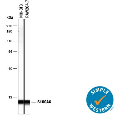
Simple Western lane view shows lysates of NIH-3T3 mouse embryonic fibroblast cell line and RAW 264.7 mouse monocyte/macrophage cell line, loaded at 0.2 mg/mL. A specific band was detected for S100A6 at approximately 7 kDa (as indicated) using 10 µg/mL of Sheep Anti-Human/Mouse S100A6 Antigen Affinity-purified Polyclonal Antibody (Catalog # AF4584) followed by 1:50 dilution of HRP-conjugated Anti-Sheep IgG Secondary Antibody (Catalog # HAF016). This experiment was conducted under reducing conditions and using the 12-230 kDa separation system.Applications for Human/Mouse S100A6 Antibody
Immunohistochemistry
Sample: Immersion fixed paraffin-embedded sections of human kidney
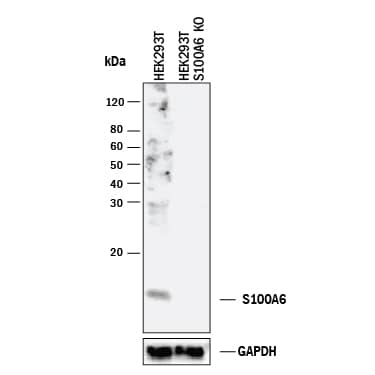
Knockout Validated
Simple Western
Sample: NIH‑3T3 mouse embryonic fibroblast cell line and RAW 264.7 mouse monocyte/macrophage cell line
Western Blot
Sample: RAW 264.7 mouse monocyte/macrophage cell line and NIH-3T3 mouse embryonic fibroblast cell line
Formulation, Preparation, and Storage
Purification
Reconstitution
Formulation
Shipping
Stability & Storage
- 12 months from date of receipt, -20 to -70 °C as supplied.
- 1 month, 2 to 8 °C under sterile conditions after reconstitution.
- 6 months, -20 to -70 °C under sterile conditions after reconstitution.
Background: S100A6
Mouse S100A6 (also prolactin receptor-associated protein and calcyclin) is a 10 kDa member of the S100 family of calcium-binding proteins. S100A6 is 89 amino acids (aa) in length and contains two calcium-binding EF-hand domains (aa 12‑47 and 48‑83). Intracellularly, S100A6 will both noncovalently homodimerize and heterodimerize with S100B plus SGT1. Extracellularly, it is secreted via a noncanonical pathway and binds to RAGE, inducing apoptosis. It is expressed by neurons, endothelium, fibroblasts and glandular epithelia. Mouse S100A6 is 99% and 96% aa identical to rat and human S100A6, respectively.
Long Name
Alternate Names
Gene Symbol
UniProt
Additional S100A6 Products
Product Documents for Human/Mouse S100A6 Antibody
Product Specific Notices for Human/Mouse S100A6 Antibody
For research use only


