Human/Mouse Semaphorin 3C Antibody
R&D Systems, part of Bio-Techne | Catalog # MAB1728

Key Product Details
Species Reactivity
Validated:
Cited:
Applications
Validated:
Cited:
Label
Antibody Source
Product Specifications
Immunogen
Gln24-Ser751 (Arg548Ala, Arg552Ala)
Accession # Q62181
Specificity
Clonality
Host
Isotype
Scientific Data Images for Human/Mouse Semaphorin 3C Antibody
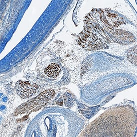
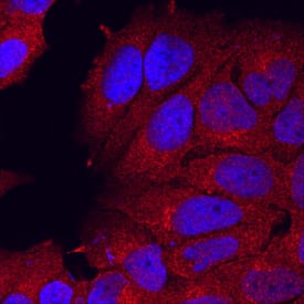
Semaphorin 3C in MCF‑7 Human Cell Line.
Semaphorin 3C was detected in immersion fixed MCF-7 human breast cancer cell line using Rat Anti-Mouse Semaphorin 3C Monoclonal Antibody (Catalog # MAB1728) at 10 µg/mL for 3 hours at room temperature. Cells were stained using the NorthernLights™ 557-conjugated Anti-Rat IgG Secondary Antibody (red; Catalog # NL013) and counterstained with DAPI (blue). Specific staining was localized to cytoplasm. View our protocol for Fluorescent ICC Staining of Cells on Coverslips.Semaphorin 3C in Mouse Embryo.
Semaphorin 3C was detected in immersion fixed frozen sections of mouse embryo (13 d.p.c.) using Rat Anti-Human/Mouse Semaphorin 3C Monoclonal Antibody (Catalog # MAB1728) at 1.7 µg/mL overnight at 4 °C. Tissue was stained using the Anti-Rat HRP-DAB Cell & Tissue Staining Kit (brown; Catalog # CTS017) and counterstained with hematoxylin (blue). Specific staining was localized to developing muscle cells. View our protocol for Chromogenic IHC Staining of Frozen Tissue Sections.Applications for Human/Mouse Semaphorin 3C Antibody
Immunocytochemistry
Sample: Immersion fixed MCF‑7 human breast cancer cell line
Immunohistochemistry
Sample: Immersion fixed frozen sections of mouse embryo (13 d.p.c.)
Western Blot
Sample: Recombinant Mouse Semaphorin 3C Fc Chimera, Truncated (Catalog # 1728-S3)
Reviewed Applications
Read 2 reviews rated 4.5 using MAB1728 in the following applications:
Formulation, Preparation, and Storage
Purification
Reconstitution
Formulation
*Small pack size (-SP) is supplied either lyophilized or as a 0.2 µm filtered solution in PBS.
Shipping
Stability & Storage
- 12 months from date of receipt, -20 to -70 °C as supplied.
- 1 month, 2 to 8 °C under sterile conditions after reconstitution.
- 6 months, -20 to -70 °C under sterile conditions after reconstitution.
Background: Semaphorin 3C
Semaphorin 3C (Sema3C; previously semaE) is one of six Class 3 secreted semaphorins which share 40-50% amino acid (aa) identity. Class 3 semaphorins are potent chemorepellents that function in axon and/or vascular guidance during development, and may be upregulated in tumor progression (1, 2). The 751 amino acid (aa) mouse Sema3C is highly modular. It contains a 20 aa signal sequence, an ~500 aa N-terminal Sema domain that forms a beta-propeller structure similar to that found in integrin molecules, a cysteine knot, a furin-type cleavage site, an Ig-like domain, and a C-terminal basic domain (1-3). Covalent dimerization plus cleavage at the C-terminus are required for activity of class 3 semaphorins (4). Mouse Sema3C shares at least 95% aa identity with human, rat, cow and dog Sema3C, and 89% and 75% aa identity with chick and zebrafish Sema3C, respectively. Type 3 semaphorins transduce signals through transmembrane plexins, either directly or by binding associated neuropilin receptors (1, 2). Sema3C signaling is transduced by Plexin-D1 indirectly via neuropilin-1 or neuropilin-2 receptors (5). Sema3C is expressed in all somitic motor neurons, in lung buds and in cardiac neural crest cells during development (1, 5-8). Sema3C activates integrins in certain cells so, in addition to its repulsive activities, it sometimes acts as a chemoattractant (6, 9). In the developing nervous system, this chemoattraction appears to complement Sema3A repulsion in adjacent cell layers (1, 6, 7). Sema3C also provides an attractive force opposing Sema6A and Sema6B to guide migration of neural crest endothelial cells to the cardiac outflow tract (10). Consequently, defects in aortic arch formation occur when Sema3C or Plexin-D1 genes or Sema3C-neuropilin interactions are disrupted (5, 11, 12).
References
- Hinck, L. (2004) Dev. Cell 7:783.
- Neufeld, G. et al. (2005) Front. Biosci. 10:751.
- Gherardi, E. et al. (2004) Curr. Opin. Struct. Biol. 14:669.
- Adams, R. H. et al. (1997) EMBO J. 16:6077.
- Gitler, A. D. et al. (2004) Dev. Cell 7:107.
- Bagnard, D. et al. (1998) Development 125:5043.
- Cohen, S. et al. (2005) Eur. J. Neurosci. 21:1767.
- Puschel, A. W. et al. (1995) Neuron 14:941.
- Herman, J. G. and G. G. Meadows (2007) Int. J. Oncol. 30:1231.
- Toyofuku, T. et al. (2008) Dev. Biol. 321:251.
- Feiner, L. et al. (2001) Development 128:3061.
- Gu, C. et al. (2003) Dev. Cell 5:45.
Alternate Names
Gene Symbol
UniProt
Additional Semaphorin 3C Products
Product Documents for Human/Mouse Semaphorin 3C Antibody
Product Specific Notices for Human/Mouse Semaphorin 3C Antibody
For research use only

