Human/Mouse TAB1 Antibody
R&D Systems, part of Bio-Techne | Catalog # AF3578

Key Product Details
Species Reactivity
Validated:
Human, Mouse
Cited:
Human
Applications
Validated:
Immunocytochemistry, Western Blot
Cited:
Immunocytochemistry, Immunohistochemistry
Label
Unconjugated
Antibody Source
Polyclonal Goat IgG
Product Specifications
Immunogen
E. coli-derived recombinant human TAB1
Ser7-Glu159
Accession # Q15750
Ser7-Glu159
Accession # Q15750
Specificity
Detects human and mouse TAB1.
Clonality
Polyclonal
Host
Goat
Isotype
IgG
Scientific Data Images for Human/Mouse TAB1 Antibody
Detection of Human/Mouse TAB1 by Western Blot.
Western blot shows lysates of WS-1 human fetal skin fibroblast cell line and C2C12 mouse myoblast cell line. PVDF membrane was probed with 1 µg/mL of Goat Anti-Human/Mouse TAB1 Antigen Affinity-purified Polyclonal Antibody (Catalog # AF3578) followed by HRP-conjugated Anti-Goat IgG Secondary Antibody (Catalog # HAF109). A specific band was detected for TAB1 at approximately 65 kDa (as indicated). This experiment was conducted under reducing conditions and using Immunoblot Buffer Group 2.TAB1 in HeLa Human Cell Line.
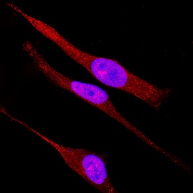
TAB1 was detected in immersion fixed HeLa human cervical epithelial carcinoma cell line using Goat Anti-Human/Mouse TAB1 Antigen Affinity-purified Polyclonal Antibody (Catalog # AF3578) at 15 µg/mL for 3 hours at room temperature. Cells were stained using the NorthernLights™ 557-conjugated Anti-Goat IgG Secondary Antibody (red; Catalog # NL001) and counterstained with DAPI (blue). Specific staining was localized to cytoplasm. View our protocol for Fluorescent ICC Staining of Cells on Coverslips.Applications for Human/Mouse TAB1 Antibody
Application
Recommended Usage
Immunocytochemistry
5-15 µg/mL
Sample: Immersion fixed human peripheral blood mononuclear cells and HeLa human cervical epithelial carcinoma cell line
Sample: Immersion fixed human peripheral blood mononuclear cells and HeLa human cervical epithelial carcinoma cell line
Western Blot
1 µg/mL
Sample: WS-1 human fetal skin fibroblast cell line and C2C12 mouse myoblast cell line
Sample: WS-1 human fetal skin fibroblast cell line and C2C12 mouse myoblast cell line
Formulation, Preparation, and Storage
Purification
Antigen Affinity-purified
Reconstitution
Reconstitute at 0.2 mg/mL in sterile PBS. For liquid material, refer to CoA for concentration.
Formulation
Lyophilized from a 0.2 μm filtered solution in PBS with Trehalose. *Small pack size (SP) is supplied either lyophilized or as a 0.2 µm filtered solution in PBS.
Shipping
Lyophilized product is shipped at ambient temperature. Liquid small pack size (-SP) is shipped with polar packs. Upon receipt, store immediately at the temperature recommended below.
Stability & Storage
Use a manual defrost freezer and avoid repeated freeze-thaw cycles.
- 12 months from date of receipt, -20 to -70 °C as supplied.
- 1 month, 2 to 8 °C under sterile conditions after reconstitution.
- 6 months, -20 to -70 °C under sterile conditions after reconstitution.
Background: TAB1
Long Name
TAK1-binding Protein
Alternate Names
MAP3K7IP1
Gene Symbol
TAB1
UniProt
Additional TAB1 Products
Product Documents for Human/Mouse TAB1 Antibody
Product Specific Notices for Human/Mouse TAB1 Antibody
For research use only
Loading...
Loading...
Loading...
Loading...

