Human/Mouse Wnt-11 Antibody
R&D Systems, part of Bio-Techne | Catalog # AF2647

Key Product Details
Validated by
Biological Validation
Species Reactivity
Validated:
Human, Mouse
Cited:
Human
Applications
Validated:
Immunocytochemistry, Immunohistochemistry, Western Blot
Cited:
Immunohistochemistry-Paraffin
Label
Unconjugated
Antibody Source
Polyclonal Goat IgG
Product Specifications
Immunogen
E. coli-derived recombinant mouse Wnt-11 peptide
Leu39-Ala79 and Lys225-Arg297
Accession # Q059Y4
Leu39-Ala79 and Lys225-Arg297
Accession # Q059Y4
Specificity
Detects mouse Wnt-11 in direct ELISAs and Western blots.
Clonality
Polyclonal
Host
Goat
Isotype
IgG
Scientific Data Images for Human/Mouse Wnt-11 Antibody
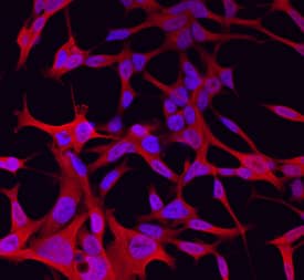
Wnt‑11 in LNCaP Human Cell Line.
Wnt-11 was detected in immersion fixed LNCaP human prostate cancer cell line using Goat Anti-Mouse Wnt-11 Antigen Affinity-purified Polyclonal Antibody (Catalog # AF2647) at 10 µg/mL for 3 hours at room temperature. Cells were stained using the NorthernLights™ 557-conjugated Anti-Goat IgG Secondary Antibody (red; Catalog # NL001) and counterstained with DAPI (blue). Specific staining was localized to cytoplasm. View our protocol for Fluorescent ICC Staining of Cells on Coverslips.Detection of Mouse Wnt-11 by Western Blot
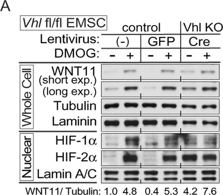
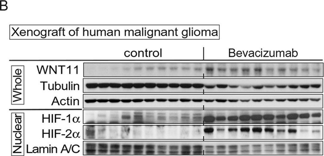
Hypoxia induces expression of WNT11 through VHL.(A,B) Higher basal levels of WNT11 protein in Vhl-deleted cells (lenti-Cre infected Vhlf/f). EMSCs isolated from Vhlf/f mouse were infected with lentivirus carrying either GFP gene (for control) or Cre recombinase (for knockout). Non-infected cells were also used as a control. Immunoblot analysis of control or Vhl KO EMSCs treated with 0.1 mM DMOG (A), and EMSCs exposed to air (21% O2) or hypoxia (1% O2) for 24 hrs (B). Laminin, alpha-tubulin, and lamin A/C were used as loading controls, WNT11 normalized to alpha-Tubulin was shown. (C,D) Inactivation of the Vhl gene results in increased Wnt11 mRNA. Wnt11 and Vegf mRNA levels in liver (C) or duodenum (D) were measured by qPCR in Liver-VhlcKO or duodenum-VhlcKO and control mice (n = 5 per group). Values normalized to Tbp mRNA are expressed relative to tissues from control mice. For panels (C,D), values are mean ± s.e.m. *p < 0.05, **p < 0.01. Image collected and cropped by CiteAb from the following publication (https://www.nature.com/articles/srep21520), licensed under a CC-BY license. Not internally tested by R&D Systems.Detection of Human Wnt-11 by Western Blot
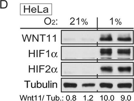
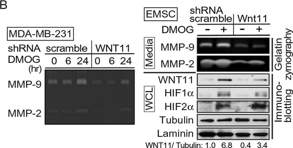
WNT11 is induced by hypoxia or hypoxic mimetics in different cell types.(A) Increased Wnt11 mRNA in EMSC adipocytes (Day 12) after hypoxia-mimetic treatments. EMSC adipocytes were treated with CoCl2 (0.1 mM), DFO (0.1 mM) or DMOG (0.1 mM) for 24 hrs. Values were normalized to Tbp mRNA and are expressed relative to control (n = 3). (B,C) Increased Wnt11 mRNA by hypoxia in EMSC preadipocytes and adipocytes (Day 0–12 after differentiation) (B), and C2C12 myoblast and myocyte (Day 0 and 8 after differentiation) (C). Wnt11 mRNA was assessed by quantitative PCR in cells exposed to air (21% O2) or hypoxia (1% O2) for 24 hrs. (n = 4). Values were normalized to Tbp mRNA and are expressed relative to 21% O2 samples (left panel). (D) Immunoblot analyses of HeLa cells under normal air or hypoxia for 24 hrs. (E,F) Induction of Wnt11 by increasing concentrations of DMOG in MDA-MB-231 cells (E) and 4T1 cells (F). (G) EMSCs treated with 0.1 mM DMOG for the indicated times. Wnt11 and Vegf mRNA expression was measured by qPCR and normalized to Tbp mRNA (n = 4). (H) WNT11 protein levels after DMOG treatment normalized to alpha-Tubulin (upper panel; n = 4). Representative immunoblots of EMSCs treated with 0.1 mM DMOG for the indicated times (Lower panel). (I) Protein expression in MDA-MB-231 cells treated with 0.1 mM DMOG. (J) Induction of Wnt11 promoter activity by hypoxia or hypoxia mimetics. pGL3-Wnt11 promoter plasmid was transfected into C2C12 cells. Cells were incubated with DMOG (left panel, n = 4) or under 21% O2 or 1% O2 (right panel, n = 8) for 24 hrs. For panels (A–C,G,H,J), values are mean ± s.e.m. *p < 0.05, **p < 0.01. For panels of immunoblotting, laminin, alpha-tubulin, and ERK were used as loading controls, WNT11 normalized to alpha-Tubulin was shown. Image collected and cropped by CiteAb from the following publication (https://www.nature.com/articles/srep21520), licensed under a CC-BY license. Not internally tested by R&D Systems.Applications for Human/Mouse Wnt-11 Antibody
Application
Recommended Usage
Immunocytochemistry
5-15 µg/mL
Sample: Immersion fixed LNCaP human prostate cancer cell line
Sample: Immersion fixed LNCaP human prostate cancer cell line
Immunohistochemistry
5-15 µg/mL
Sample: Immersion fixed frozen sections of mouse embryo (E13-15)
Sample: Immersion fixed frozen sections of mouse embryo (E13-15)
Western Blot
0.1 µg/mL
Sample: Recombinant Mouse Wnt-11
Sample: Recombinant Mouse Wnt-11
Formulation, Preparation, and Storage
Purification
Antigen Affinity-purified
Reconstitution
Reconstitute at 0.2 mg/mL in sterile PBS. For liquid material, refer to CoA for concentration.
Formulation
Lyophilized from a 0.2 μm filtered solution in PBS with Trehalose. *Small pack size (SP) is supplied either lyophilized or as a 0.2 µm filtered solution in PBS.
Shipping
Lyophilized product is shipped at ambient temperature. Liquid small pack size (-SP) is shipped with polar packs. Upon receipt, store immediately at the temperature recommended below.
Stability & Storage
Use a manual defrost freezer and avoid repeated freeze-thaw cycles.
- 12 months from date of receipt, -20 to -70 °C as supplied.
- 1 month, 2 to 8 °C under sterile conditions after reconstitution.
- 6 months, -20 to -70 °C under sterile conditions after reconstitution.
Background: Wnt-11
Long Name
Wingless-type MMTV Integration Site Family, Member 11
Alternate Names
Wnt11
Gene Symbol
WNT11
UniProt
Additional Wnt-11 Products
Product Documents for Human/Mouse Wnt-11 Antibody
Product Specific Notices for Human/Mouse Wnt-11 Antibody
For research use only
Loading...
Loading...
Loading...
Loading...
