Human/Mouse Wnt-5a Antibody
R&D Systems, part of Bio-Techne | Catalog # MAB645

Key Product Details
Species Reactivity
Validated:
Cited:
Applications
Validated:
Cited:
Label
Antibody Source
Product Specifications
Immunogen
Gln38-Lys380
Accession # P22725
Specificity
Clonality
Host
Isotype
Scientific Data Images for Human/Mouse Wnt-5a Antibody
Wnt‑5a in Mouse Embryo.
Wnt-5a was detected in immersion fixed frozen sections of mouse embryo using Human/Mouse Wnt-5a Monoclonal Antibody (Catalog # MAB645) at 10 µg/mL overnight at 4 °C. Tissue was stained using the NorthernLights™ 557-conjugated Anti-Rat IgG Secondary Antibody (orange; Catalog # NL013) and counter-stained with DAPI (blue). View our protocol for Fluorescent IHC Staining of Frozen Tissue Sections.Detection of Mouse Wnt-5a by Immunocytochemistry/Immunofluorescence
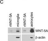
WNT-5A expression in mouse GFAP+astrocytes. (A) Immunohistochemistry was performed on adult mouse brain sections using an anti-WNT-5A antibody in combination with anti-glial fibrillary acidic protein (GFAP) and ionized calcium-binding adaptor protein1 (IBA1) antibodies as astrocyte and microglia marker, respectively. Merge presents the overlay of IBA1, GFAP, WNT-5A. Size bar −2 μm. The images represent a maximum intensity projection of a Z-stack of 5 μm thickness. The white square marked ‘B’ indicates the area magnified in B: (B) Close up of a GFAP+ astrocyte reveals the expression of WNT-5A in this cell type. (C) shows immunoblot detection of recombinant WNT-5A (rWNT-5A; 375 ng/lane) in comparison to lysates from mouse primary microglia and mixed astrocyte cultures. beta-actin serves as a loading control. (D) The bar graph depicts expression levels of WNT-5A mRNA in mouse primary microglia and mixed astrocyte cultures measured by QPCR. Data are normalized to GAPDH expression and analyzed with a non-parametric Mann–Whitney test. *, P < 0.05; **, P < 0.01; ***, P < 0.001. n = 4 to 8. (E) shows indirect immunocytochemistry of mixed astrocyte cultures employing anti-GFAP as astrocyte and anti-CD11b as microglia markers. DAPI is used as nuclear counterstain. Image represents a maximum image projection of an 8 μm Z-stack. Size bar 20 μm. The frame shows 63 cells in total and 10 CD11b-positive microglia (arrows). Routinely 10% to 18% microglia were observed (n = 4). DAPI, 4',6-diamidino-2-phenylindole; GADPH, glyceraldehyde 3-phosphate dehydrogenase; n, number. Image collected and cropped by CiteAb from the following publication (https://pubmed.ncbi.nlm.nih.gov/22647544), licensed under a CC-BY license. Not internally tested by R&D Systems.Detection of Mouse Wnt-5a by Western Blot
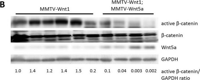
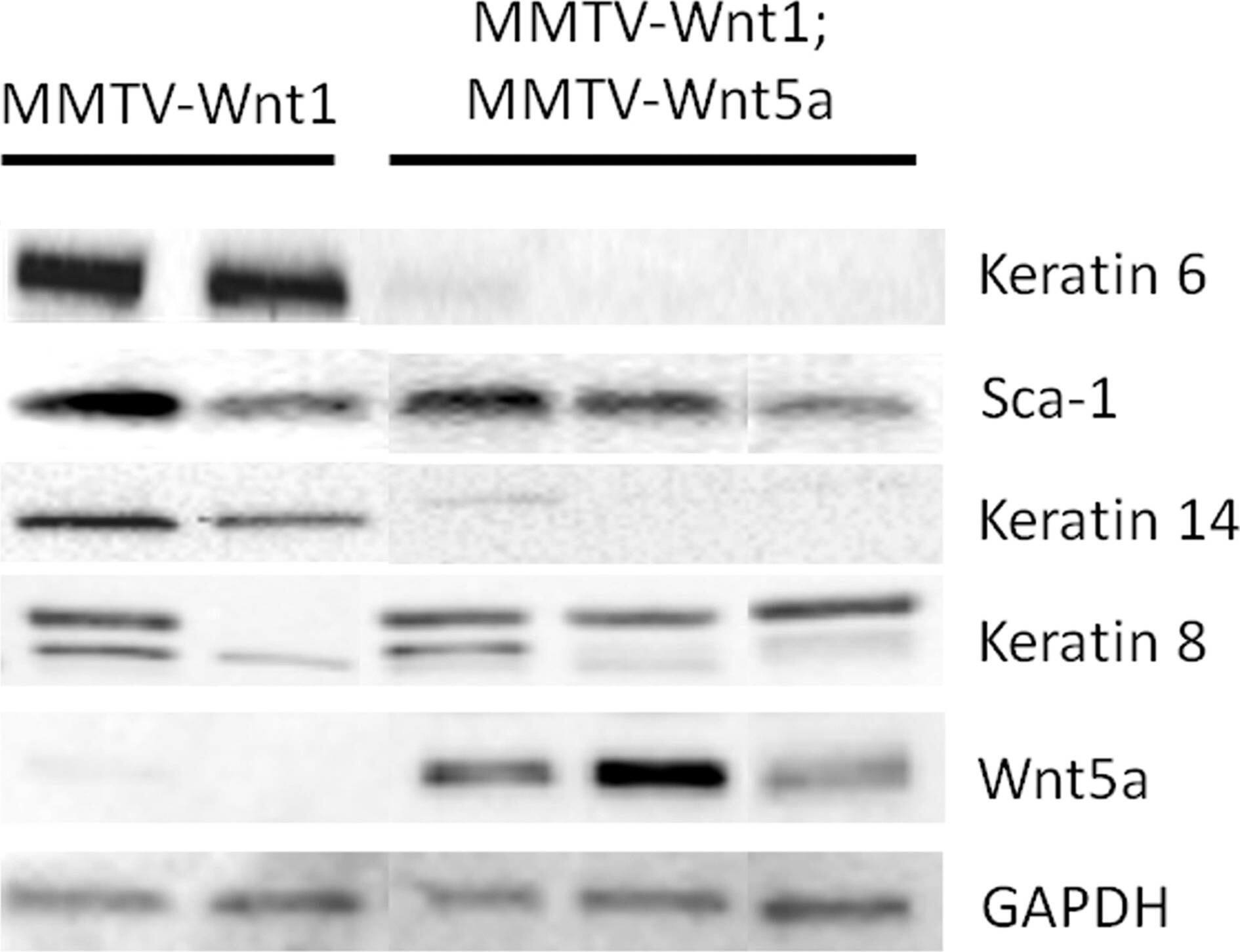
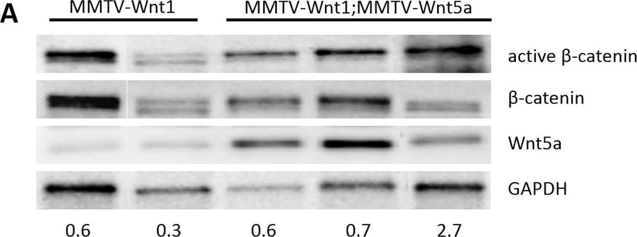
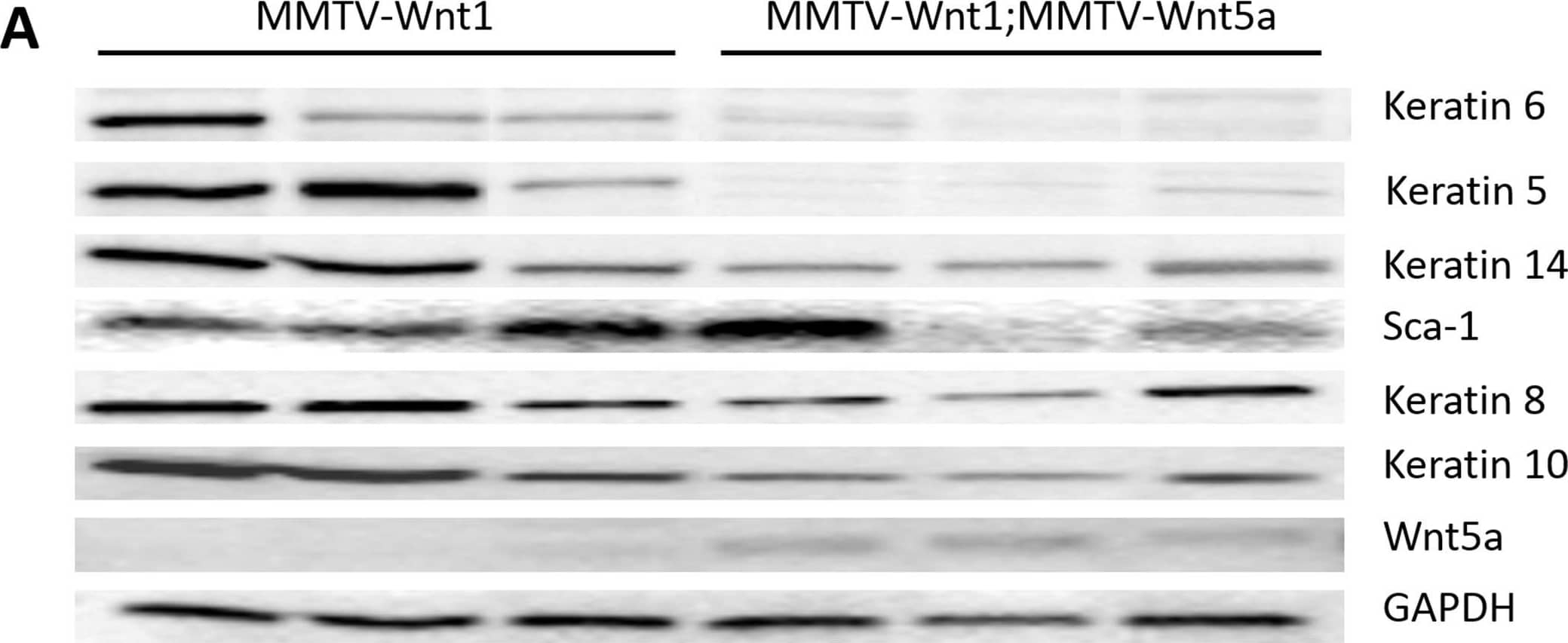
Wnt5a expressing tumors have less Wnt/ beta-catenin signaling than MMTV-Wnt1 tumors.(A) Quantitative RT-PCR of Wnt/ beta-catenin target genes. Expression of Axin2 mRNA in MMTV-Wnt1 versus MMTV-Wnt1;MMTV-Wnt5a tumors as determined by quantitative RT-PCR (n = 5 MMTV-Wnt1, n = 5 MMTV-Wnt1;MMTV-Wnt5a). Data are shown as tables obtained using REST software. Axin2 mRNA was significantly down-regulated in MMTV-Wnt1;MMTV-Wnt5a tumors. (B) Western blot for beta-catenin protein. Protein lysates were prepared from MMTV-Wnt1 and MMTV-Wnt1;MMTV-Wnt5a tumors. beta-catenin and glyceraldehyde 3-phosphate dehydrogenase (GAPDH) were used as loading controls. The ratio of active beta-catenin to beta-catenin as determined by densitometic analysis is shown. MMTV-Wnt1;MMTV-Wnt5a tumors displayed decreased levels of active beta-catenin compared to controls. Image collected and cropped by CiteAb from the following publication (https://dx.plos.org/10.1371/journal.pone.0113247), licensed under a CC-BY license. Not internally tested by R&D Systems.Applications for Human/Mouse Wnt-5a Antibody
Immunohistochemistry
Sample: Immersion-fixed frozen sections of mouse embryo
Reviewed Applications
Read 2 reviews rated 4 using MAB645 in the following applications:
Formulation, Preparation, and Storage
Purification
Reconstitution
Formulation
Shipping
Stability & Storage
- 12 months from date of receipt, -20 to -70 °C as supplied.
- 1 month, 2 to 8 °C under sterile conditions after reconstitution.
- 6 months, -20 to -70 °C under sterile conditions after reconstitution.
Background: Wnt-5a
Wnt proteins are secreted glycoproteins that contain a conserved pattern of 23‑24 cysteine residues. Wnts play critical roles in both carcinogenesis and embryonic development for a variety of organisms. Wnts bind to receptors of the Frizzled family, sometimes in conjunction with other membrane-associated proteins such as LRPs or proteoglycans. Downstream effects of Wnt signaling occur through different intracellular components, depending on which pathway is activated. Three pathways have been characterized: the canonical Wnt/ beta-catenin pathway, the Wnt/Ca2+ pathway, and the planar cell polarity (1‑2).
Wnt-5a is part of the subgroup of Wnts that are not axis-inducing in Xenopus embryos and do not transform C57MG mammary epithelial cells. This subgroup is also implicated in the Wnt/Ca2+ pathway, playing roles in cell movements and cell adhesion (3). This non-canonical Wnt pathway can inhibit canonical Wnt/ beta-catenin signaling. In Wnt-5a deficient mouse embryos, beta-catenin accumulates in the limb bud suggesting that Wnt-5a normally promotes degradation of beta-catenin (4). Likewise, in Xenopus embryos Wnt-5a antagonizes the ability of the canonical Wnt subgroup to induce a secondary axis (5). Wnt-5a is implicated in various types of cancer and has complex roles. It acts as a tumor suppressor for mammary, B-cell, colon, and uroepithelial cancer cells but is up-regulated in melanomas, where expression levels correlate with severity of metastasis (3). Furthermore, aberrant Wnt-5a signaling results in other diseases such as rheumatoid arthritis (6). Like other developmental growth factors Wnt-5a has diverse roles in development. They are too numerous to enunciate here, as functions span from early anterior-posterior development and gastrulation movements to maintaining hematopoietic stem cell population, lung morphogenesis, and limb outgrowth. Mature Wnt-5a is a 49 kDa protein that shares 99% amino acid identity in mouse, rat and human.
References
- Miller, J.R. (2002) Genome Biol. 3:3001.
- Roelink, H. and R. Nusse (1991) Genes Dev. 5:381.
- Veeman, M.T. et al. (2003) Developmental Cell 5:367.
- Topol, L. et al. (2003) J. Cell Biol 162:899.
- Torres, M. et al. (1996) J. Cell Biol. 133:1123.
- Sen, M. et al. (2001) Arthritis & Rheumatism 44:772.
Long Name
Alternate Names
Gene Symbol
UniProt
Additional Wnt-5a Products
Product Documents for Human/Mouse Wnt-5a Antibody
Product Specific Notices for Human/Mouse Wnt-5a Antibody
For research use only

