Human NG2/MCSP Antibody
R&D Systems, part of Bio-Techne | Catalog # AF2585

Key Product Details
Validated by
Species Reactivity
Validated:
Cited:
Applications
Validated:
Cited:
Label
Antibody Source
Product Specifications
Immunogen
Ser1583-Ser2224
Accession # Q6UVK1
Specificity
Clonality
Host
Isotype
Scientific Data Images for Human NG2/MCSP Antibody
Detection of Human NG2/MCSP by Western Blot.
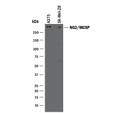
Western blot shows lysates of A375 human melanoma cell line and SK-Mel-28 human malignant melanoma cell line. PVDF membrane was probed with 0.5 µg/mL of Goat Anti-Human NG2/MCSP Antigen Affinity-purified Polyclonal Antibody (Catalog # AF2585) followed by HRP-conjugated Anti-Goat IgG Secondary Antibody (Catalog # HAF017). A specific band was detected for NG2/MCSP at approximately 300 kDa (as indicated). This experiment was conducted under reducing conditions and using Immunoblot Buffer Group 1.Western Blot Shows Human NG2/MCSP Specificity by Using Knockout Cell Line.
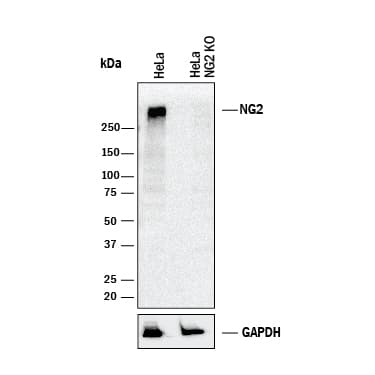
Western blot shows lysates of HeLa human cervical epithelial carcinoma parental cell line and NG2/MCSP knockout HeLa cell line (KO). PVDF membrane was probed with 0.5 µg/mL of Goat Anti-Human NG2/MCSP Antigen Affinity-purified Polyclonal Antibody (Catalog # AF2585) followed by HRP-conjugated Anti-Goat IgG Secondary Antibody (Catalog # HAF017). A specific band was detected for NG2/MCSP at approximately 300 kDa (as indicated) in the parental HeLa cell line, but is not detectable in knockout HeLa cell line. GAPDH (Catalog # AF5718) is shown as a loading control. This experiment was conducted under reducing conditions and using Immunoblot Buffer Group 1.Applications for Human NG2/MCSP Antibody
Knockout Validated
Western Blot
Sample: A375 human melanoma cell line and SK‑Mel‑28 human malignant melanoma cell line
Formulation, Preparation, and Storage
Purification
Reconstitution
Formulation
Shipping
Stability & Storage
- 12 months from date of receipt, -20 to -70 °C as supplied.
- 1 month, 2 to 8 °C under sterile conditions after reconstitution.
- 6 months, -20 to -70 °C under sterile conditions after reconstitution.
Background: NG2/MCSP
NG2 (neuron/glia-type 2 antigen; also MCSP/melanoma chondroitin sulfate proteoglycan) is a 250‑500 kDa type I integral membrane proteoglycan found on synantocytes (NG2+ glia), vascular pericytes, chondroblasts, macrophages and melanoma cells. It binds multiple components of the ECM, and serves as a ligand for alpha3 beta1 and alpha4 beta1 integrins. Mature human NG2 is 2293 amino acids (aa) in length. It contains a large 2195 aa extracellular region (aa 30‑2224) and a short 79 aa cytoplasmic tail. NG2 is variably glycanated. Without proteoglycan, NG2 is expressed as a 250 kDa glycoprotein. Juxtamembrane proteolysis generates a soluble ECD that noncovalently associates with the transmembrane fragment. Over aa 1583‑2224, human NG2 is 81% aa identical to both mouse and canine NG2.
Long Name
Alternate Names
Gene Symbol
UniProt
Additional NG2/MCSP Products
Product Documents for Human NG2/MCSP Antibody
Product Specific Notices for Human NG2/MCSP Antibody
For research use only

