Human NGFR/TNFRSF16 Antibody
R&D Systems, part of Bio-Techne | Catalog # MAB3671

Key Product Details
Species Reactivity
Applications
Label
Antibody Source
Product Specifications
Immunogen
Lys29-Asn250
Accession # P08138
Specificity
Clonality
Host
Isotype
Scientific Data Images for Human NGFR/TNFRSF16 Antibody
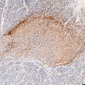
NGFR/TNFRSF16 in Human Spinal Cord Tissue.
NGFR/TNFRSF16 was detected in immersion fixed paraffin-embedded sections of human spinal cord tissue using Mouse Anti-Human NGFR/TNFRSF16 Monoclonal Antibody (Catalog # MAB3671) at 5 µg/mL for 1 hour at room temperature followed by incubation with the Anti-Mouse IgG VisUCyte™ HRP Polymer Antibody (VC001). Before incubation with the primary antibody, tissue was subjected to heat-induced epitope retrieval using Antigen Retrieval Reagent-Basic (CTS013). Tissue was stained using DAB (brown) and counterstained with hematoxylin (blue). Specific staining was localized to dorsal horn. Staining was performed using IHC Staining with VisUCyte HRP Polymer Detection Reagents protocol.Applications for Human NGFR/TNFRSF16 Antibody
Immunohistochemistry
Sample: Immersion fixed paraffin-embedded sections of human spinal cord tissue
Formulation, Preparation, and Storage
Purification
Reconstitution
Formulation
Shipping
Stability & Storage
- 12 months from date of receipt, -20 to -70 °C as supplied.
- 1 month, 2 to 8 °C under sterile conditions after reconstitution.
- 6 months, -20 to -70 °C under sterile conditions after reconstitution.
Background: NGFR/TNFRSF16
p75 neurotrophin receptor, also named low affinity NGF receptor (NGF R), is a type I transmembrane protein that belongs to the tumor necrosis factor receptor family. NGF R cDNA encodes a 427 amino acid (aa) residue precursor protein with a 28 aa residue signal peptide, a 222 aa residue extracellular domain, a 22 aa residue transmembrane domain and a 155 aa residue intracellular domain. The extracellular region contains four cysteine-rich domains and binds NGF, BDNF, NT-3 and NT-4 approximately equally with low affinity. The cytoplasmic region contains a subtype 2 death domain.
NGF R expression has been shown to occur widely during development and in the adult. Expression has been detected in both neuronal and non-neuronal cells. NGF R was originally reported to function as a positive regulator of TrkA activity. NGF R has also been shown to signal by itself. Depending on its cellular environment, NGF R has now been shown to regulate cell migration, gene expression and to mediate apoptosis. Recombinant NGF R/Fc chimera binds NGF with high affinity and is a potent NGF antagonist. Naturally occurring truncated NGF R containing the extracellular domain and lacking the transmembrane or intracellular domain has been detected in vivo in urine, plasma and in amniotic fluid of humans and rats.
References
- Barker, P.A. and R.A. Murphy (1992) Molecular and Cellular Biochemistry 110:1.
- Bamji, A.X. et al. (1998) J. Cell Biol. 140:911.
- Feinstein, E. et al. (1995) Trends Biochem. Sci. 20:342.
Long Name
Alternate Names
Gene Symbol
UniProt
Additional NGFR/TNFRSF16 Products
Product Documents for Human NGFR/TNFRSF16 Antibody
Product Specific Notices for Human NGFR/TNFRSF16 Antibody
For research use only
