Human NGL-1/LRRC4C Antibody
R&D Systems, part of Bio-Techne | Catalog # MAB4899

Key Product Details
Species Reactivity
Applications
Label
Antibody Source
Product Specifications
Immunogen
Gln45-Lys527 (predicted)
Accession # Q9HCJ2
Specificity
Clonality
Host
Isotype
Scientific Data Images for Human NGL-1/LRRC4C Antibody
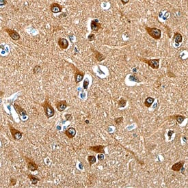
NGL‑1/LRRC4C in Human Spinal Cord.
NGL-1/LRRC4C was detected in immersion fixed paraffin-embedded sections of human spinal cord using Mouse Anti-Human NGL-1/LRRC4C Monoclonal Antibody (Catalog # MAB4899) at 15 µg/mL overnight at 4 °C. Tissue was stained using the Anti-Mouse HRP-DAB Cell & Tissue Staining Kit (brown; Catalog # CTS002) and counter-stained with hematoxylin (blue). Specific staining was localized to the dorsal horn. View our protocol for Chromogenic IHC Staining of Paraffin-embedded Tissue Sections.Applications for Human NGL-1/LRRC4C Antibody
Immunohistochemistry
Sample: Immersion fixed paraffin-embedded sections of human spinal cord
Formulation, Preparation, and Storage
Purification
Reconstitution
Formulation
Shipping
Stability & Storage
- 12 months from date of receipt, -20 to -70 °C as supplied.
- 1 month, 2 to 8 °C under sterile conditions after reconstitution.
- 6 months, -20 to -70 °C under sterile conditions after reconstitution.
Background: NGL-1/LRRC4C
Human NGL-1 (Netrin-G1 ligand; gene name LRRC4C) is a 67 kDa (predicted for mature protein), type I transmembrane cell adhesion molecule that is a member of the NGL family of proteins (1, 2). It is synthesized from a precursor that is 640 amino acids (aa) in length that contains a 44 aa signal sequence, a 483 aa extracellular region, a 21 aa transmembrane region, and a short cytoplasmic tail of 92 aa. The extracellular region of NGL-1 consists of nine leucine-rich repeats (LRRs) that are flanked by LRR N-terminal and LRR C-terminal domains, and followed by an Ig-like C2‑type domain (1, 2). The cytoplasmic region contains a C‑terminal Glu-Thr-Gln-Ile sequence that corresponds to a potential PDZ (postsynaptic density-95/discs large/zona occludens-1) domain-binding motif (1, 2). Human NGL-1 shares 99.7% aa sequence identity with mouse NGL-1. Mouse NGL-1 is highly expressed in the developing cerebral cortex and the striatum at embryonic day 14 (1). Postnatally, NGL-1 is expressed exclusively in the brain, with the highest expression found in the cerebral cortex as a whole, and in individual neocortical areas such as the frontal, parietal and occipital lobes (1). Moderate expression of NGL-1 occurs in the putamen, amygdala, hippocampus and medulla oblongata (1). Weak expression is found in the caudate nucleus and thalamus (1). Functionally, membrane-bound cell-surface NGL-1 binds to netrin-G1 specifically through its LRR region, and in the developing brain, may promote neurite outgrowth of thalamocortical axons (1‑4).
References
- Lin, J.C. et al. (2003) Nat. Neurosci. 6:1270.
- Kim, S. et al. (2006) Nat. Neurosci. 9:1294.
- Chen, Y. et al. (2006) Brain Res. Rev. 51:265.
- Nishimura-Akiyoshi, S. et al. (2007) Proc. Natl. Acad. Sci. U.S.A. 104:14801.
Long Name
Alternate Names
Gene Symbol
UniProt
Additional NGL-1/LRRC4C Products
Product Documents for Human NGL-1/LRRC4C Antibody
Product Specific Notices for Human NGL-1/LRRC4C Antibody
For research use only
