Human NTB-A/SLAMF6 APC-conjugated Antibody
R&D Systems, part of Bio-Techne | Catalog # FAB19081A


Key Product Details
Species Reactivity
Validated:
Cited:
Applications
Validated:
Cited:
Label
Antibody Source
Product Specifications
Immunogen
Leu28-Lys225
Accession # Q96DU3
Specificity
Clonality
Host
Isotype
Scientific Data Images for Human NTB-A/SLAMF6 APC-conjugated Antibody
Detection of NTB-A/SLAMF6 in Human Blood Lymphocytes by Flow Cytometry.
Human peripheral blood lymphocytes were stained with Mouse Anti-Human NTB-A/SLAMF6 APC-conjugated Monoclonal Antibody (Catalog # FAB19081A, filled histogram) or isotype control antibody (Catalog # IC003A, open histogram). View our protocol for Staining Membrane-associated Proteins.Detection of Human NTB-A/SLAMF6/CD352 by Flow Cytometry
Functional activity of Rev1-Vpu expressed from pCG expression plasmids.(A) CMV-IE promoter-based pCG expression vectors containing vpu (left panel) or the rev1-vpu fusion gene (right panel). An enhanced version of the green fluorescent protein (eGFP) is co-expressed via an IRES. (B) Expression of Rev1-Vpu and Vpu in HEK293T cells transfected with the indicated pCG vectors. A Vpu-specific antiserum was used for detection. eGFP was detected to check transfection efficiencies. (C-F) FACS analysis of (C) CD4, (D) tetherin, (E) CD1d or (F) NTB-A receptor modulation by ZM247 Vpu and Rev1-Vpu. HEK293T cells were transfected with expression vectors for the respective surface receptor and Vpu or Rev1-Vpu. 40 h post transfection, surface receptor levels were monitored by two-color flow cytometry. Dot plots indicating the gating strategy are shown in the right panels. Bar diagrams summarizing three to five independent experiments +/- SD are shown on the left (***p<0.001; **p<0.01; *p<0.05; n.s. not significant). Image collected and cropped by CiteAb from the following publication (https://dx.plos.org/10.1371/journal.pone.0142118), licensed under a CC-BY license. Not internally tested by R&D Systems.Detection of Human NTB-A/SLAMF6/CD352 by Flow Cytometry
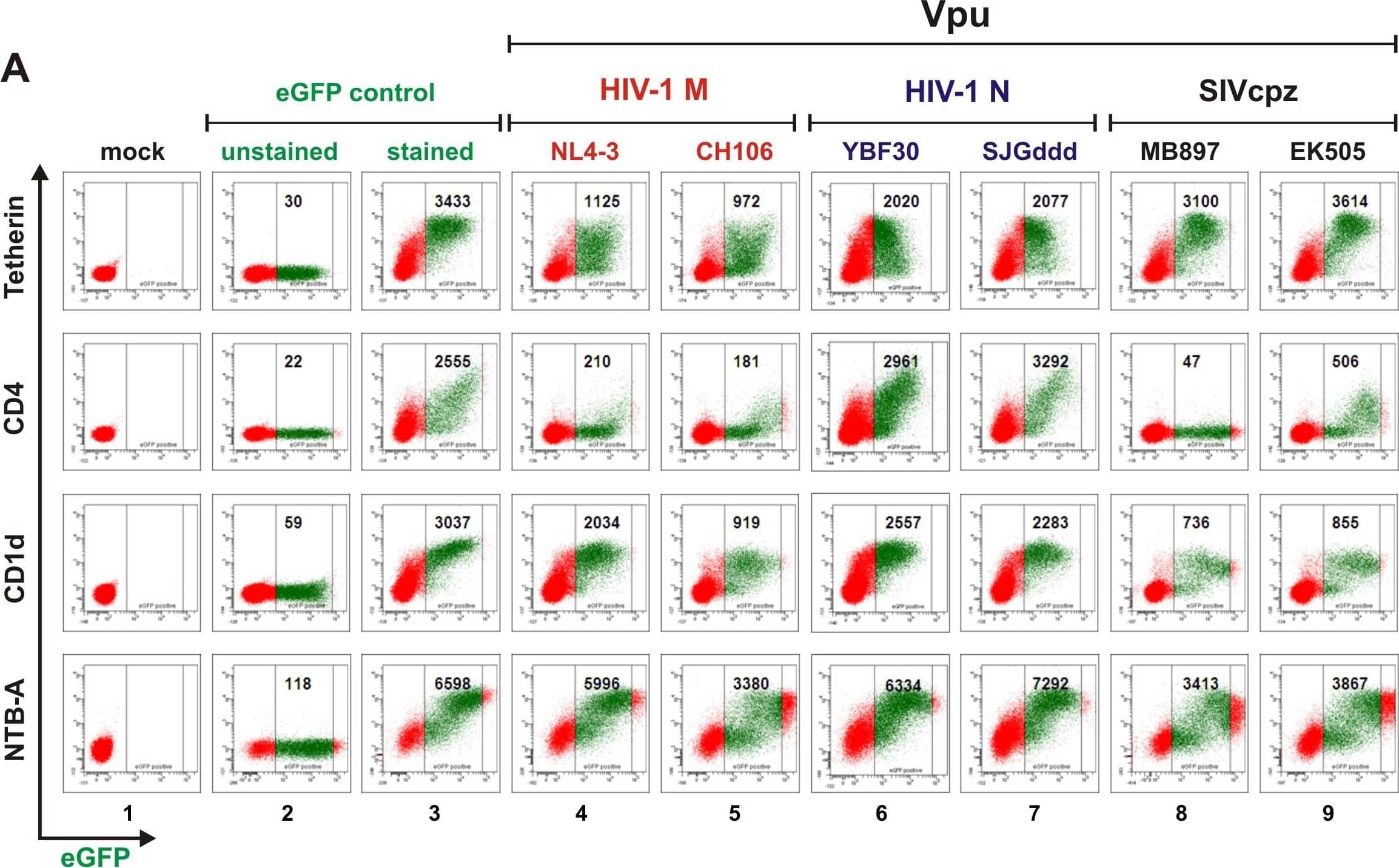
Functional characterization of Vpus from Cameroonian HIV-1 N strains.(A) FACS analysis of 293T cells cotransfected with tetherin, CD4, CD1d or NTB-A expression vectors and pCGCG plasmids expressing eGFP alone (lanes 2 and 3) or together with the indicated vpu allele (lanes 4 to 9). eGFP expression levels used to calculate receptor downmodulation and the mean fluorescence intensities (MFIs) are indicated. (B) Effect of various Vpus on infectious virus release. 293T cells were cotransfected with HIV-1 deltaVpu NL4-3 (2 µg), pCGCG vectors coexpressing eGFP and Vpu (500 ng), and a construct expressing HU tetherin (125 ng). Viral supernatants were obtained two days later and used to measure the quantity of infectious HIV-1 in the culture supernatants by infecting TZM-bl indicator cells. Shown is the infectious virion yield relative to that obtained in the absence of tetherin (100%). The results were confirmed in two independent experiments and each symbol indicates infectious virus yield in the presence of one of the 25 vpu alleles analyzed. (C–F) Vpu-dependent reduction of (C) tetherin, (D) CD4, (E) CD1d and (F) NTB-A surface expression in 293T cells. Shown is the reduction in the levels of receptor cell surface expression relative to those measured in cells transfected with the eGFP only control vector. Each symbol represents n-fold downmodulation of the indicated receptor molecule by one individual vpu allele examined. Shown are average values derived from three experiments. Vpu alleles derived from HIV-1 M are color coded red, N blue (except for the DJO0131 Vpu highlighted in green) and SIVcpz black; those shown in panel A are indicated by open symbols in panels B to F. Image collected and cropped by CiteAb from the following open publication (https://pubmed.ncbi.nlm.nih.gov/23308067), licensed under a CC-BY license. Not internally tested by R&D Systems.Applications for Human NTB-A/SLAMF6 APC-conjugated Antibody
Flow Cytometry
Sample: Human peripheral blood lymphocytes
Formulation, Preparation, and Storage
Purification
Formulation
Shipping
Stability & Storage
- 12 months from date of receipt, 2 to 8 °C as supplied.
Background: NTB-A/SLAMF6
NTB-A (NK-T-B-antigen), also known as Ly108 and SLAMF6, is a 60 kDa type I transmembrane glycoprotein that belongs to the SLAM subgroup of the CD2 family (1). Mature human NTB-A contains a 205 amino acid (aa) extracellular domain (ECD) with one Ig-like V-set and one Ig-like C2-set domain. It also contains a 21 aa transmembrane segment and an 84 aa cytoplasmic domain with two immunoreceptor tyrosine-based switch motifs (ITSMs) (2, 3). An alternately spliced isoform is truncated in the cytoplasmic domain and lacks the two ITSMs. Within the ECD, human NTB-A shares 48% aa sequence identity with mouse and rat NTB-A. The ECD of human NTB-A shares 19%‑34% aa sequence identity with comparable regions of human 2B4, BLAME, CD2F-10, CD84, CD229, CRACC, and SLAM. NTB-A is expressed on the surface of NK, T, and B lymphocytes as well as eosinophils (2, 4, 5). It interacts homophilically through weak associations between the Ig-V domains (2, 5‑7). NTB-A functions as an activating coreceptor on NK and T cells (2, 5, 6, 8). Tyrosine phosphorylation in the membrane proximal ITSM enables specific association with EAT-2, an interaction that is required for NTB-A mediated cytotoxicity of NK cells (9). Phosphorylation-dependent NTB-A association with SAP is required for full production of IFN-gamma by NK cells (5, 9). This interaction is independent of EAT-2 binding and appears to involve the membrane distal ITSM (5, 9). NTB-A deficient mice show weakened Th2 responses and elevated levels of neutrophil-derived inflammatory mediators (10). On B cells, NTB-A modulates immunoglobulin class switching and the balance between tolerance and autoimmunity (5, 11).
References
- Veillette, A. (2006) Immunol. Rev. 214:22.
- Bottino, C. et al. (2001) J. Exp. Med. 194:235.
- Fraser, C.C. et al. (2002) Immunogenetics 53:843.
- Munitz, A. et al. (2005) J. Immunol. 174:110.
- Valdez, P.A. et al. (2004) J. Biol. Chem. 279:18662.
- Flaig, R.M. et al. (2004) J. Immunol. 172:6524.
- Cao, E. et al. (2006) Immunity 25:559.
- Stark, S. and C. Watzl (2006) Int. Immunol. 18:241.
- Eissmann, P. and C. Watzl (2006) J. Immunol. 177:3170.
- Howie, D. et al. (2005) J. Immunol. 174:5931.
- Kumar, K.R. et al. (2006) Science 312:1665.
Long Name
Alternate Names
Entrez Gene IDs
Gene Symbol
UniProt
Additional NTB-A/SLAMF6 Products
Product Documents for Human NTB-A/SLAMF6 APC-conjugated Antibody
Product Specific Notices for Human NTB-A/SLAMF6 APC-conjugated Antibody
For research use only

