Human Osteoactivin/GPNMB Antibody
R&D Systems, part of Bio-Techne | Catalog # AF2550

Key Product Details
Validated by
Species Reactivity
Validated:
Cited:
Applications
Validated:
Cited:
Label
Antibody Source
Product Specifications
Immunogen
Lys23-Asn486
Accession # NP_002501
Specificity
Clonality
Host
Isotype
Scientific Data Images for Human Osteoactivin/GPNMB Antibody
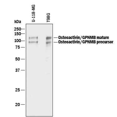
Detection of Human Osteoactivin/GPNMB by Western Blot.
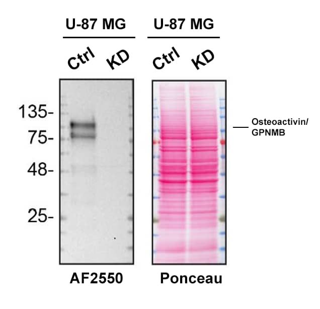
Western blot shows lysates of U-118-MG human glioblastoma/astrocytoma cell line and T98G human glioblastoma cell line. PVDF membrane was probed with 0.5 µg/mL of Goat Anti-Human Osteoactivin/GPNMB Antigen Affinity-purified Polyclonal Antibody (Catalog # AF2550) followed by HRP-conjugated Anti-Goat IgG Secondary Antibody (HAF017). Specific bands were detected for Osteoactivin/GPNMB at approximately 95 and 120 kDa (as indicated). This experiment was conducted under reducing conditions and using Immunoblot Buffer Group 1.Osteoactivin/GPNMB in Human Liver.
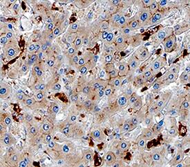
Osteoactivin/GPNMB was detected in immersion fixed paraffin-embedded sections of human liver using Goat Anti-Human Osteoactivin/GPNMB Antigen Affinity-purified Polyclonal Antibody (Catalog # AF2550) at 3 µg/mL overnight at 4 °C. Tissue was stained using the Anti-Goat HRP-DAB Cell & Tissue Staining Kit (brown; CTS008) and counterstained with hematoxylin (blue). Specific staining was localized to Kupffer cells. View our protocol for Chromogenic IHC Staining of Paraffin-embedded Tissue Sections.Osteoactivin/GPNMB Specificity is Shown by Immunocytochemistry in Knockdown Cell Line.
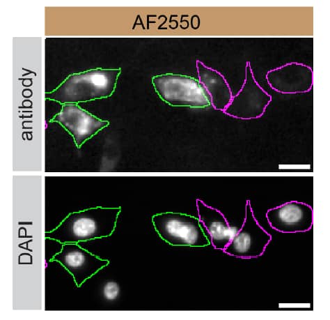
U-87 MG human glioblastoma/astrocytoma parental cell line ctrl and Osteoactivin/GPNMB U-87 MG KD cells were labelled with a green or a far-red fluorescent dye, respectively. Cells were stained with Goat Anti-Human Osteoactivin/GPNMB Antigen Affinity-purified Polyclonal Antibody (Catalog # AF2550) followed by incubation with an Alexa-fluor 555 conjugated secondary antibody (upper panel). DAPI-only counterstained cells shown on a lower panel. Acquisition of the blue (nucleus-DAPI), green (identification of ctrl cells), red (antibody staining) and far-red (identification of KD cells) channels was performed. Representative images of the blue and red (grayscale) channels are shown. Ctrl and KD cells are outlined with green and magenta dashed line, respectively. Primary antibody concentration used: 0.2 µg/mL. Image, protocol and testing courtesy of YCharOS Inc. (ycharos.com).Applications for Human Osteoactivin/GPNMB Antibody
Immunohistochemistry
Sample: Immersion fixed paraffin-embedded sections of human liver
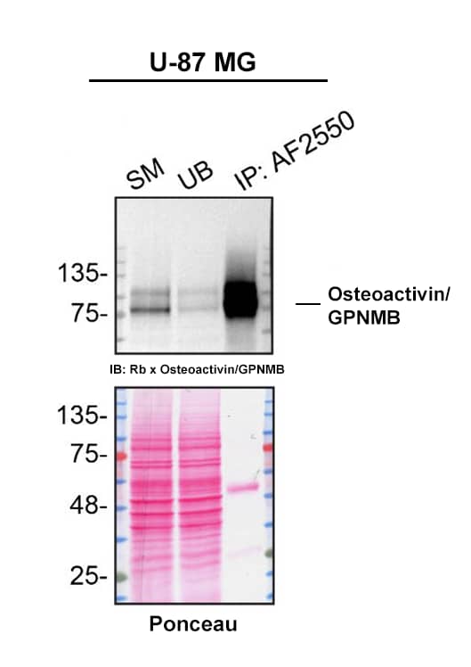
Immunoprecipitation
Sample: Cell lysate of U-87 MG human glioblastoma/astrocytoma cell line
Western Blot
Sample: U‑118‑MG human glioblastoma/astrocytoma cell line and T98G human glioblastoma cell line
Human Osteoactivin/GPNMB Sandwich Immunoassay
Reviewed Applications
Read 3 reviews rated 5 using AF2550 in the following applications:
Formulation, Preparation, and Storage
Purification
Reconstitution
Formulation
*Small pack size (-SP) is supplied either lyophilized or as a 0.2 µm filtered solution in PBS.
Shipping
Stability & Storage
- 12 months from date of receipt, -20 to -70 °C as supplied.
- 1 month, 2 to 8 °C under sterile conditions after reconstitution.
- 6 months, -20 to -70 °C under sterile conditions after reconstitution.
Background: Osteoactivin/GPNMB
Osteoactivin (also GPNMB and DC-HIL) is a variably glycosylated 75 - 125 kDa member of the NMB/pMEL-17 family of molecules. It is found in multiple subcellular sites, but is most often associated with the endosomal/lysosomal compartment (1-3). Human Osteoactivin is a 560 amino acid (aa) type I transmembrane protein. Its precursor contains a 21 aa signal sequence, a 465 aa luminal/extracellular domain, a 21 aa transmembrane segment and a 53 aa cytoplasmic tail (4, 5). The luminal region contains an N-terminal heparin-binding motif (aa 23-26), multiple glycosylation sites, an RGD motif (aa 64-66) and an 88 aa PKD domain (aa 240-327). The intracellular tail has an ITAM (Y-x-x-I) and lysosomal targeting (L-L) motif (4, 5). The extracellular/luminal region shares 74% and 77% aa identity with the equivalent regions in mouse and canine, respectively. Multiple isoforms would appear to exist. There is one alternate splice form known that shows a 12 aa insert between aa 339-340 (6). An addtional 206 aa isoform shows a mutation at position # 181 that results in a 26 aa substitution for the C-terminal 380 amino acids (7, 8). This has the potential to be soluble and may represent a counterpart to a secreted isoform of rat Osteoactivin (9). Cells known to express Osteoactivin include macrophages/Kupffer cells, fibroblasts, osteoblasts, myeloid dendritic cells, retinal pigment epithelial cells and melanocytes, plus fetal chondrocytes and stratum basale keratinocytes (3-5, 10-12). In mice, Osteoactivin is reported to bind to heparan sulfate-proteoglycan, possibly on the surface of endothelial cells and may also interact with integrins (13). It also appears to act as an inflammatory suppressor gene, as its expression downregulates the macrophage inflammatory response by inhibiting IL-6 and IL-12 p40 production (3).
References
- Bachner, D. et al. (2002) Gene Exp. Patterns 1:159.
- Safadi, F.F. et al. (2002) J. Cell. Biochem. 84:12.
- Ripoll, V.M. et al. (2007) J. Immunol. 178:6557.
- Owen, T.A. et al. (2003) Crit. Rev. Eukaryot. Gene Expr. 13:205.
- Weterman, M.A.J. et al. (1995) Int. J. Cancer 60:73.
- Kuan, C-T. et al. (2006) Clin. Cancer Res. 12:1970.
- Lennerz, V. et al. (2005) Proc. Natl. Acad. Sci. USA 102:16013.
- Genbank Accession # AAH11595.
- Abdelmagid, S.M. et al. (2007) J. Cell. Physiol. 210:26.
- Haralanova-Ilieva, B. et al. (2005) J. Hepatol. 42:565.
- Ahn, J.H. et al. (2002) Blood 100:1742.
- Anderson, M.G. et al. (2002) Nat. Genet. 30:81.
- Shikano, S. et al. (2001) J. Biol. Chem. 276:8125.
Long Name
Alternate Names
Gene Symbol
UniProt
Additional Osteoactivin/GPNMB Products
Product Documents for Human Osteoactivin/GPNMB Antibody
Product Specific Notices for Human Osteoactivin/GPNMB Antibody
For research use only
