Human Otolin-1 Antibody
R&D Systems, part of Bio-Techne | Catalog # MAB8045

Key Product Details
Species Reactivity
Applications
Label
Antibody Source
Product Specifications
Immunogen
Lys24-Pro477
Accession # A6NHN0
Specificity
Clonality
Host
Isotype
Scientific Data Images for Human Otolin-1 Antibody
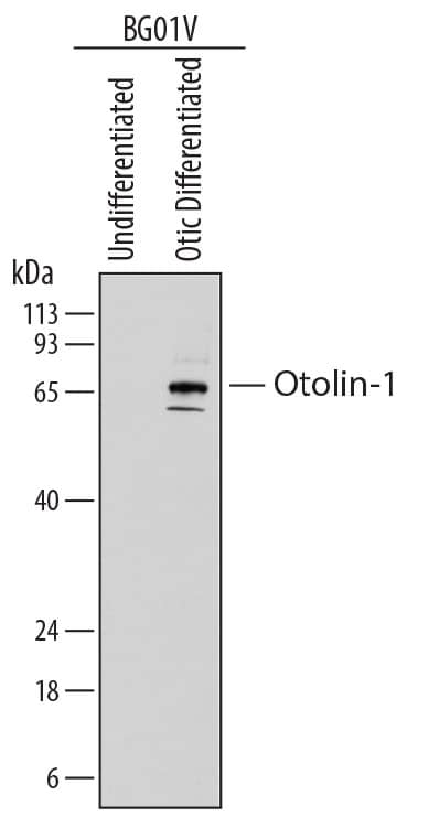
Detection of Human Otolin-1 by Western Blot.
Western blot shows lysates of BG01V human embryonic stem cells undifferentiated or differentiated to early otic lineage. PVDF membrane was probed with 0.25 µg/mL of Mouse Anti-Human Otolin-1 Monoclonal Antibody (Catalog # MAB8045) followed by HRP-conjugated Anti-Mouse IgG Secondary Antibody (Catalog # HAF018). Specific bands were detected for Otolin-1 at approximately 60-70 kDa (as indicated). This experiment was conducted under reducing conditions and using Immunoblot Buffer Group 1.Otolin-1 in differentiated BG01V Human Embryonic Stem Cells.
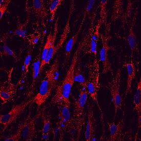
Otolin-1 was detected in immersion fixed BG01V human embryonic stem cells differentiated to early otic lineage using Mouse Anti-Human Otolin-1 Monoclonal Antibody (Catalog # MAB8045) at 10 µg/mL for 3 hours at room temperature. Cells were stained using the NorthernLights™ 557-conjugated Anti-Mouse IgG Secondary Antibody (red; Catalog # NL007) and counterstained with DAPI (blue). Specific staining was localized to cytoplasm. View our protocol for Fluorescent ICC Staining of Cells on Coverslips.Applications for Human Otolin-1 Antibody
Immunocytochemistry
Sample: Immersion fixed BG01V human embryonic stem cells differentiated to early otic lineage
Western Blot
Sample: BG01V human embryonic stem cells differentiated to early otic lineage
Formulation, Preparation, and Storage
Purification
Reconstitution
Formulation
Shipping
Stability & Storage
- 12 months from date of receipt, -20 to -70 °C as supplied.
- 1 month, 2 to 8 °C under sterile conditions after reconstitution.
- 6 months, -20 to -70 °C under sterile conditions after reconstitution.
Background: Otolin-1
Otolin (OTOL1), also known as C1qTNF15, is an approximately 65 kDa protein found in the otoconial membrane lining the cochlea and vestibular labyrinth of the inner ear. It is secreted by supporting cells of the sensory epithelium. The otoconial membrane contains particles known as otoconia which are composed of glycoproteins and proteoglycans coated with calcium carbonate crystals. Otolin is one of the protein components of otoconia particles, and it is important for otoconia formation as well as for auditory and vestibular function. It associates into multimers and disulfide-linked oligomers and also associates with other otoconial proteins including Cerebellin-1 and Otoconin-90. Otolin contains three collagen-like regions followed by a C1q-like domain at the C-terminus. It is extensively glycosylated and has multiple hydroxylated proline residues in the collagenous regions. Human Otolin shares 72% aa sequence identity with mouse and rat Otolin.
Alternate Names
Gene Symbol
UniProt
Additional Otolin-1 Products
Product Documents for Human Otolin-1 Antibody
Product Specific Notices for Human Otolin-1 Antibody
For research use only

