Human p67phox Antibody
R&D Systems, part of Bio-Techne | Catalog # AF7830

Key Product Details
Species Reactivity
Applications
Label
Antibody Source
Product Specifications
Immunogen
Lys355-Val526 (His389Gln)
Accession # P19878
Specificity
Clonality
Host
Isotype
Scientific Data Images for Human p67phox Antibody
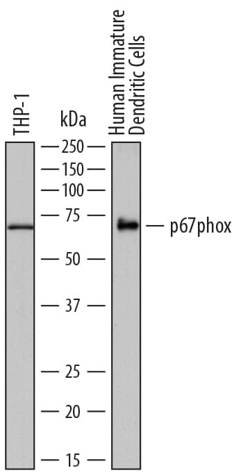
Detection of Human p67phox by Western Blot.
Western blot shows lysates of THP-1 human acute monocytic leukemia cell line and human immature dendritic cells. PVDF membrane was probed with 0.5 µg/mL of Sheep Anti-Human p67phox Antigen Affinity-purified Polyclonal Antibody (Catalog # AF7830) followed by HRP-conjugated Anti-Sheep IgG Secondary Antibody (Catalog # HAF016). A specific band was detected for p67phox at approximately 70 kDa (as indicated). This experiment was conducted under reducing conditions and using Immunoblot Buffer Group 1.Applications for Human p67phox Antibody
Western Blot
Sample: THP‑1 human acute monocytic leukemia cell line and human immature dendritic cells
Formulation, Preparation, and Storage
Purification
Reconstitution
Formulation
Shipping
Stability & Storage
- 12 months from date of receipt, -20 to -70 °C as supplied.
- 1 month, 2 to 8 °C under sterile conditions after reconstitution.
- 6 months, -20 to -70 °C under sterile conditions after reconstitution.
Background: p67phox
P67phox (67 kDa phagocyte oxidase; also NCF2 and NADPH oxidase activator 2) is a 67-70 kDa cytosolic member of the NCF2/NOXA1 family of proteins. It is expressed in multiple cell types, including B cells, endothelial cells, phagocytic cells such as neutrophils and macrophages and, to some degree, fibroblasts. Upon phagocytosis, phagocytes generate superoxide for the purpose of killing ingested microbes. This occurs through the formation and activation of a complex termed NADPH oxidase, which consists of membrane-associated cytochrome b558, plus Rac GTPase and three SH3 domain-containing components, p47phox and a p67phox:p40-phox dimer. Human p67phox is 526 amino acids (aa) in length. It contains two TPR repeats that participate in scaffold formation (aa 11-98), an SH3 domain (aa 240-299), an OPR motif that regulates PB1 interactions (aa 351-429), and a C-terminal SH3 domain (aa 457-516). There is one splice variant that shows a deletion of aa 231-293, and multiple point mutations along the length of the molecule. Over aa 355-526, human p67phox shares 76% aa identity with mouse p67phox.
Alternate Names
Gene Symbol
UniProt
Additional p67phox Products
Product Documents for Human p67phox Antibody
Product Specific Notices for Human p67phox Antibody
For research use only
