Human PLD2 Antibody
R&D Systems, part of Bio-Techne | Catalog # AF10123

Key Product Details
Species Reactivity
Applications
Label
Antibody Source
Product Specifications
Immunogen
Met1-His117
Accession # O14939
Specificity
Clonality
Host
Isotype
Scientific Data Images for Human PLD2 Antibody
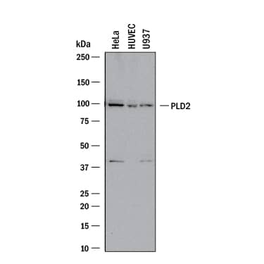
Detection of Human PLD2 by Western Blot.
Western blot shows lysates of HeLa human cervical epithelial carcinoma cell line, HUVEC human umbilical vein endothelial cells, and U937 human histiocytic lymphoma cell line. PVDF membrane was probed with 1 µg/mL of Goat Anti-Human PLD2 Antigen Affinity-purified Polyclonal Antibody (Catalog # AF10123) followed by HRP-conjugated Anti-Goat IgG Secondary Antibody (Catalog # HAF017). A specific band was detected for PLD2 at approximately 95 kDa (as indicated). This experiment was conducted under reducing conditions and using Immunoblot Buffer Group 1.PLD2 in Human Colon Cancer Tissue.
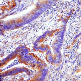
PLD2 was detected in immersion fixed paraffin-embedded sections of human colon cancer tissue using Goat Anti-Human PLD2 Antigen Affinity-purified Polyclonal Antibody (Catalog # AF10123) at 10 µg/mL for 1 hour at room temperature followed by incubation with the Anti-Goat IgG VisUCyte™ HRP Polymer Antibody (Catalog # VC004). Before incubation with the primary antibody, tissue was subjected to heat-induced epitope retrieval using Antigen Retrieval Reagent-Basic (Catalog # CTS013). Tissue was stained using DAB (brown) and counterstained with hematoxylin (blue). Specific staining was localized to cytoplasm in epithelial cells. View our protocol for IHC Staining with VisUCyte HRP Polymer Detection Reagents.Applications for Human PLD2 Antibody
Immunohistochemistry
Sample: Immersion fixed paraffin-embedded sections of human colon cancer tissue
Western Blot
Sample: HeLa human cervical epithelial carcinoma cell line, HUVEC human umbilical vein endothelial cells, and U937 human histiocytic lymphoma cell line
Formulation, Preparation, and Storage
Purification
Reconstitution
Formulation
Shipping
Stability & Storage
- 12 months from date of receipt, -20 to -70 °C as supplied.
- 1 month, 2 to 8 °C under sterile conditions after reconstitution.
- 6 months, -20 to -70 °C under sterile conditions after reconstitution.
Background: PLD2
Phospholipase D2 (PLD2) is an enzyme encoded by the PLD2 gene. Phosphatidylcholine (PC)-specific phospholipases D (PLDs) catalyze the hydrolysis of PC to produce phosphatidic acid and choline. The Phospholipase D (PLD) lipid-signaling enzyme superfamily has long been studied for its roles in cell communication and a wide range of cell biological processes. Potent PLD2 inhibitors are available, like VU 0364739 hydrochloride (Tocris Cat. 4171) and ML 298 hydrochloride (Tocris Cat. 4895). Three different PLD2 isoforms -PLD2A, PLD2B and PLD2C- have been described, ranging from 38 to 105 KDa.
Long Name
Alternate Names
Gene Symbol
UniProt
Additional PLD2 Products
Product Documents for Human PLD2 Antibody
Product Specific Notices for Human PLD2 Antibody
For research use only

