Human RIPK2/RIP2 Antibody
R&D Systems, part of Bio-Techne | Catalog # MAB103871

Key Product Details
Species Reactivity
Applications
Label
Antibody Source
Product Specifications
Immunogen
Met1-Met540
Accession # O43353
Specificity
Clonality
Host
Isotype
Scientific Data Images for Human RIPK2/RIP2 Antibody
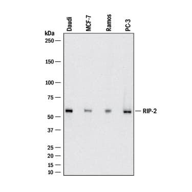
Detection of Human RIPK2/RIP2 by Western Blot.
Western blot shows lysates of Daudi human Burkitt's lymphoma cell line, MCF-7 human breast cancer cell line, Ramos human Burkitt's lymphoma cell line, and PC-3 human prostate cancer cell line. PVDF membrane was probed with 2 µg/mL of Mouse Anti-Human RIPK2/RIP2 Monoclonal Antibody (Catalog # MAB103871) followed by HRP-conjugated Anti-Mouse IgG Secondary Antibody (Catalog # HAF018). A specific band was detected for RIPK2/RIP2 at approximately 62 kDa (as indicated). This experiment was conducted under reducing conditions and using Immunoblot Buffer Group 1.RIPK2/RIP2 in Human Tonsil.
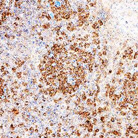
RIPK2/RIP2 was detected in immersion fixed paraffin-embedded sections of human tonsil using Mouse Anti-Human RIPK2/RIP2 Monoclonal Antibody (Catalog # MAB103871) at 5 µg/mL for 1 hour at room temperature followed by incubation with the Anti-Mouse IgG VisUCyte™ HRP Polymer Antibody (Catalog # VC001). Before incubation with the primary antibody, tissue was subjected to heat-induced epitope retrieval using Antigen Retrieval Reagent-Basic (Catalog # CTS013). Tissue was stained using DAB (brown) and counterstained with hematoxylin (blue). Specific staining was localized to lymphocytes. View our protocol for IHC Staining with VisUCyte HRP Polymer Detection Reagents.Detection of Human RIPK2/RIP2 by Simple WesternTM.
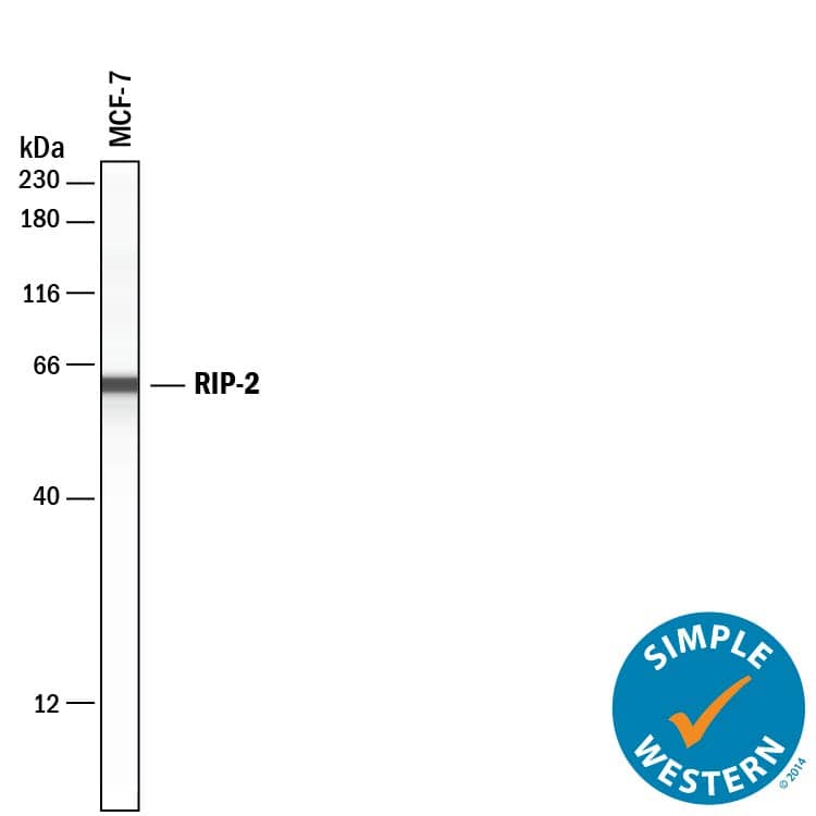
Simple Western lane view shows lysates of MCF‑7 human breast cancer cell line, loaded at 0.2 mg/mL. A specific band was detected for RIPK2/RIP2 at approximately 62 kDa (as indicated) using 20 µg/mL of Mouse Anti-Human RIPK2/RIP2 Monoclonal Antibody (Catalog # MAB103871) . This experiment was conducted under reducing conditions and using the 12-230 kDa separation system.Applications for Human RIPK2/RIP2 Antibody
Immunohistochemistry
Sample: Immersion fixed paraffin-embedded sections of human tonsil
Simple Western
Sample: MCF‑7 human breast cancer cell line
Western Blot
Sample: Daudi human Burkitt's lymphoma cell line, MCF‑7 human breast cancer cell line, Ramos human Burkitt's lymphoma cell line, and PC‑3 human prostate cancer cell line
Formulation, Preparation, and Storage
Purification
Reconstitution
Formulation
Shipping
Stability & Storage
- 12 months from date of receipt, -20 to -70 °C as supplied.
- 1 month, 2 to 8 °C under sterile conditions after reconstitution.
- 6 months, -20 to -70 °C under sterile conditions after reconstitution.
Background: RIPK2/RIP2
Receptor-interacting serine/threonine-protein kinase 2, also known as RIPK2 or RIP-2, is a 540 amino acids (aa) Serine/threonine/tyrosine kinase encoded by the RIPK2 gene. RIP-2, a member of the receptor-interacting protein (RIP) family of serine/threonine protein kinases, plays an essential role in modulation of innate and adaptive immune responses. Human RIP2 is an adapter protein necessary for signal propagation of the Nucleotide-binding-oligomerization-domain-containing proteins 1/2 (NOD1 and NOD2). Upon stimulation by bacterial peptidoglycans, NOD1 and NOD2 are activated, oligomerize, and recruit RIPK2 through the C-terminal caspase recruitment domain (CARD). Human and mouse RIPK2 aa sequences are 84% identical .
Long Name
Alternate Names
Gene Symbol
UniProt
Additional RIPK2/RIP2 Products
Product Documents for Human RIPK2/RIP2 Antibody
Product Specific Notices for Human RIPK2/RIP2 Antibody
For research use only


