Detection of Human SCGF/CLEC11a by Western Blot
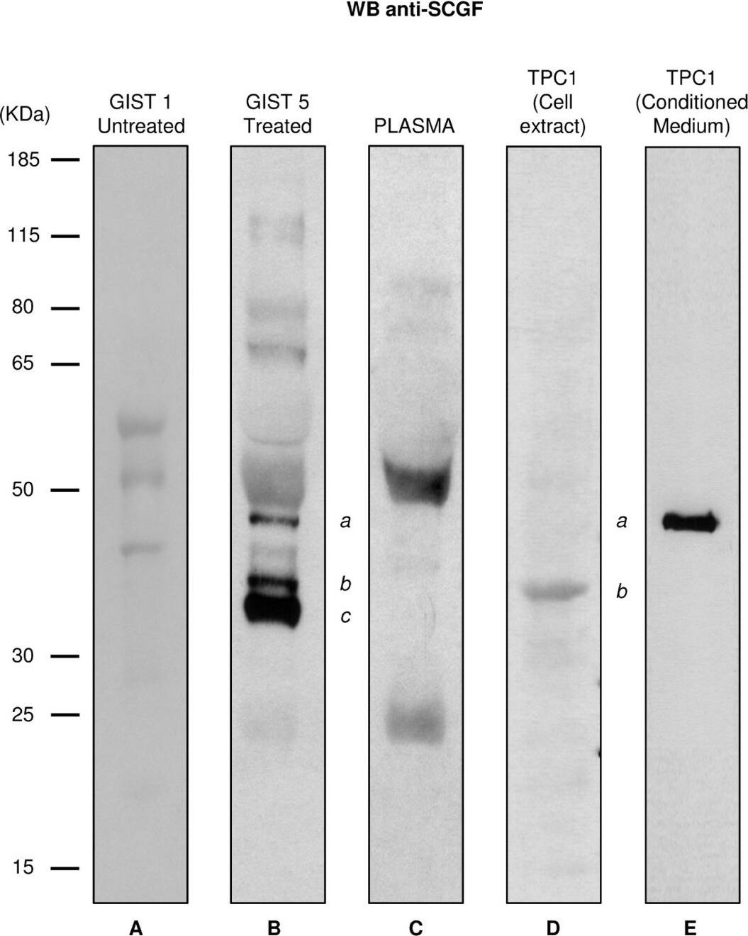
Western blotting of SCGF expression. Western blotting with anti SCGF-alpha /-beta antibody of tumor tissue samples from the untreated GIST 1 patient (lane A), the SCGF-positive GIST 5 patient (lane B), a pool of plasma samples from healthy subjects (lane C), and the cell extract (lane D) and conditioned medium (lane E) from the TPC1 cell line. (a) indicates the mature and secreted form of SCGF-alpha detected in the GIST 5 sample (lane B) and in the TPC1 conditioned medium (lane E); (b) indicates the immature and cytoplasmatic form of SCGF-alpha detected in the GIST 5 sample (lane B; corresponding to band 8 in Figure 1A) and in the TPC1 cell extract (lane D); (c) points out the quantitatively most relevant form, exclusively present in the GIST 5 sample, corresponding to band 9 in Figure 1A. Image collected and cropped by CiteAb from the following publication (https://translational-medicine.biomedcentral.com/articles/10.1186/1479-5876-9-158), licensed under a CC-BY license. Not internally tested by R&D Systems.
Detection of Human SCGF/CLEC11a by Western Blot
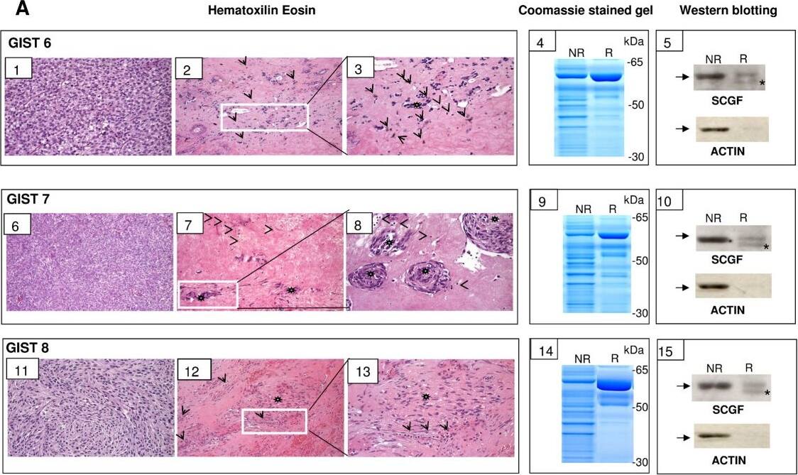
Analysis of an independent set of imatinib-treated GIST samples. A) Comparative analysis of tumoral areas from of GIST 6, 7 and 8, which resulted non-responding or responding to imatinib treatment. Panels 1-5, 6-10 and 11-15 refer to GISTs 6, 7 and 8 respectively. Panels 1, 6 and 11 represent non-responding areas; panels 2, 7 and 12 responding areas; panels 3, 8 and 13 show the inset areas of panels 2, 7, and 12. Arrows and arrowheads indicate inflammatory macrophages and lymphocytes, respectively, and asterisks residual tumor cells. Panels 4, 9 and 14 show the protein lysates prepared from adjacent tumor areas were separated onto SDS gels and stained with Coomassie-blu. Lane NR indicates the non-responding area and lane R indicates the responding area. Panels 5, 10 and 15 show NR and R protein lysates were probed with anti-SCGF and anti-actin antibodies, respectively. Note that in panels 5, 10 and 15 arrows refer to the actin band that is detected as a non-specific background band in anti-SCGF blot. As expected, due to the higher cellularity we observe a more intense actin band in NR samples than in R samples. B) Western blotting of SCGF expression in GIST 9-16 samples. GIST 9-13 represent protein lysates from non-responder GIST samples and GIST 14-16 protein lysates from responder samples. C corresponds to GIST 6 protein lysate. Samples were probed with both anti-SCGF and anti-actin antibodies. Arrow indicates actin band and asterisks SCGF band. In non-responder samples anti-SCGF detected exclusively the actin band. Image collected and cropped by CiteAb from the following publication (https://translational-medicine.biomedcentral.com/articles/10.1186/1479-5876-9-158), licensed under a CC-BY license. Not internally tested by R&D Systems.
Detection of Human SCGF/CLEC11a by Immunohistochemistry
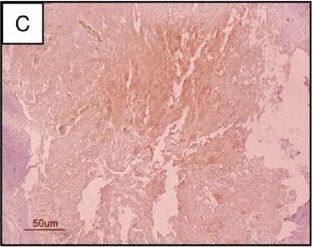
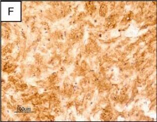
Immunohistochemistry of CD68 and SCGF in GIST 5 and GIST 2. A) Hematoxilin-eosin section of one representative slide from GIST 5. B) CD68 staining; CD68 positive macrophages are scattered through the section. C) SCGF staining; SCGF positivity was observed in the corresponding stromal area. D) Hematoxylin-eosin section of one representative slide from GIST 2. E) CD68 staining; a diffuse infiltration of CD68-positive macrophages is visible in this area. F) SCGF staining; SCGF positivity was observed in the corresponding stromal counterpart. Image collected and cropped by CiteAb from the following publication (https://translational-medicine.biomedcentral.com/articles/10.1186/1479-5876-9-158), licensed under a CC-BY license. Not internally tested by R&D Systems.
Detection of Human SCGF/CLEC11a by Immunohistochemistry
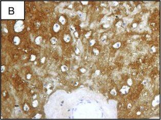
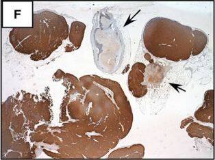
Immunohistochemistry of SCGF expression in GIST samples. A) Hematoxylin-eosin section of one representative slide from GIST 5. B) SCGF staining; strong SCGF positivity, consistent with western blotting, was restricted to the stroma compartment. C) CD117 staining; cells retained weak cytoplasmic KIT reactivity. The rare viable tumoral cells present in the responding area were SCGF-negative in the cytoplasm and the nucleus. D) Hematoxylin-eosin section of one representative slide from GIST 2. Highly cellulated areas indicating the absence of pathological response are visible. Only two areas showing rare tumoral cells are present. E) CD117 staining; all cellulated areas exhibit strong KIT expression not observed in the acellulated areas mostly composed of stroma. F) SCGF staining; strong SCGF expression is restricted to the non-cellulated areas, in contrast to CD117 staining. Image collected and cropped by CiteAb from the following publication (https://translational-medicine.biomedcentral.com/articles/10.1186/1479-5876-9-158), licensed under a CC-BY license. Not internally tested by R&D Systems.
Detection of Human SCGF/CLEC11a by Western Blot
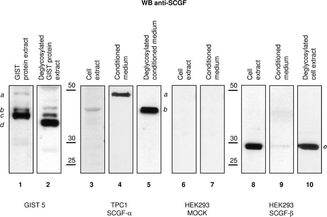
Western blotting of SCGF after deglycosylation. Western blotting with anti-SCGF-alpha /-beta antibody. Lanes 1 and 2 compare SCGF patterns in the untreated (lane 1) and the deglycosylated (lane 2) protein extract of the GIST 5 sample. Lanes 3-5 contain a comparison of the cytoplasmatic (lane 3) and the secreted (lane 4) SCGF-alpha forms and the untreated (lane 4) and the deglycosylated secreted (lane 5) SCGF-alpha forms in the TPC1 cell line. Lanes 6 and 7 depict the expression of SCGF-alpha and -beta in the cell extract and conditioned medium of a mock HEK293 cell line (negative control). Lanes 8-10 reflect SCGF-beta expression in the cell extract (lane 8) and conditioned medium (lane 9) of the transfected HEK293 cell line; lanes 8 and 10 show the untreated and the deglycosylated SCGF-beta forms in the transfected HEK293 cell line extract. The mature and secreted forms of SCGF-alpha detected in the GIST 5 sample (lane 1) and the TPC1 conditioned medium (lane 4) are indicated by (a); (b) indicates an immature, cytoplasmatic, unglycosylated form of SCGF-alpha in the untreated (lane 1) and deglycosylated (lane 2) GIST 5 samples, and in the cell extract (lane 3) and conditioned medium of the TPC1 cell line after deglycosylation (lane 5); the form indicated by (c) was exclusively detected in the GIST 5 sample, was the quantitatively most relevant form in the untreated sample (lane 1), and was observed after deglycosylation (lane 2); (d) indicates the form exclusively detected and quantitatively most relevant in the deglycosylated GIST 5 sample (lane 2), corresponding to the protein backbone of SCGF-alpha (~36 kDa); (e) indicates the form detected in the untreated (lane 8) and deglycosylated (lane 10) cell extract of the transfected HEK293 cell line, corresponding to the primary structure of SCGF-beta (~27 kDa). Image collected and cropped by CiteAb from the following publication (https://translational-medicine.biomedcentral.com/articles/10.1186/1479-5876-9-158), licensed under a CC-BY license. Not internally tested by R&D Systems.
Detection of Human Human SCGF/CLEC11a Antibody by Western Blot
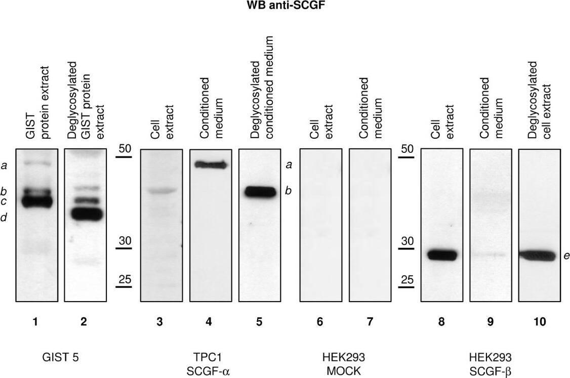
Western blotting of SCGF after deglycosylation. Western blotting with anti-SCGF-alpha /-beta antibody. Lanes 1 and 2 compare SCGF patterns in the untreated (lane 1) and the deglycosylated (lane 2) protein extract of the GIST 5 sample. Lanes 3-5 contain a comparison of the cytoplasmatic (lane 3) and the secreted (lane 4) SCGF-alpha forms and the untreated (lane 4) and the deglycosylated secreted (lane 5) SCGF-alpha forms in the TPC1 cell line. Lanes 6 and 7 depict the expression of SCGF-alpha and -beta in the cell extract and conditioned medium of a mock HEK293 cell line (negative control). Lanes 8-10 reflect SCGF-beta expression in the cell extract (lane 8) and conditioned medium (lane 9) of the transfected HEK293 cell line; lanes 8 and 10 show the untreated and the deglycosylated SCGF-beta forms in the transfected HEK293 cell line extract. The mature and secreted forms of SCGF-alpha detected in the GIST 5 sample (lane 1) and the TPC1 conditioned medium (lane 4) are indicated by (a); (b) indicates an immature, cytoplasmatic, unglycosylated form of SCGF-alpha in the untreated (lane 1) and deglycosylated (lane 2) GIST 5 samples, and in the cell extract (lane 3) and conditioned medium of the TPC1 cell line after deglycosylation (lane 5); the form indicated by (c) was exclusively detected in the GIST 5 sample, was the quantitatively most relevant form in the untreated sample (lane 1), and was observed after deglycosylation (lane 2); (d) indicates the form exclusively detected and quantitatively most relevant in the deglycosylated GIST 5 sample (lane 2), corresponding to the protein backbone of SCGF-alpha (~36 kDa); (e) indicates the form detected in the untreated (lane 8) and deglycosylated (lane 10) cell extract of the transfected HEK293 cell line, corresponding to the primary structure of SCGF-beta (~27 kDa). Image collected and cropped by CiteAb from the following publication (https://pubmed.ncbi.nlm.nih.gov/21943129), licensed under a CC-BY license. Not internally tested by R&D Systems.
Detection of Human Human SCGF/CLEC11a Antibody by Western Blot
Analysis of an independent set of imatinib-treated GIST samples. A) Comparative analysis of tumoral areas from of GIST 6, 7 and 8, which resulted non-responding or responding to imatinib treatment. Panels 1-5, 6-10 and 11-15 refer to GISTs 6, 7 and 8 respectively. Panels 1, 6 and 11 represent non-responding areas; panels 2, 7 and 12 responding areas; panels 3, 8 and 13 show the inset areas of panels 2, 7, and 12. Arrows and arrowheads indicate inflammatory macrophages and lymphocytes, respectively, and asterisks residual tumor cells. Panels 4, 9 and 14 show the protein lysates prepared from adjacent tumor areas were separated onto SDS gels and stained with Coomassie-blu. Lane NR indicates the non-responding area and lane R indicates the responding area. Panels 5, 10 and 15 show NR and R protein lysates were probed with anti-SCGF and anti-actin antibodies, respectively. Note that in panels 5, 10 and 15 arrows refer to the actin band that is detected as a non-specific background band in anti-SCGF blot. As expected, due to the higher cellularity we observe a more intense actin band in NR samples than in R samples. B) Western blotting of SCGF expression in GIST 9-16 samples. GIST 9-13 represent protein lysates from non-responder GIST samples and GIST 14-16 protein lysates from responder samples. C corresponds to GIST 6 protein lysate. Samples were probed with both anti-SCGF and anti-actin antibodies. Arrow indicates actin band and asterisks SCGF band. In non-responder samples anti-SCGF detected exclusively the actin band. Image collected and cropped by CiteAb from the following publication (https://pubmed.ncbi.nlm.nih.gov/21943129), licensed under a CC-BY license. Not internally tested by R&D Systems.


