Human SPINK1 Antibody
R&D Systems, part of Bio-Techne | Catalog # AF7496

Key Product Details
Species Reactivity
Applications
Label
Antibody Source
Product Specifications
Immunogen
Asp24-Cys79
Accession # P00995
Specificity
Clonality
Host
Isotype
Scientific Data Images for Human SPINK1 Antibody
Detection of Human SPINK1 by Western Blot.
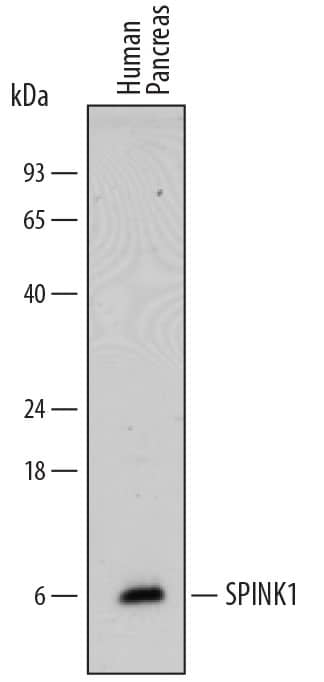
Western blot shows lysates of human pancreas tissue. PVDF membrane was probed with 0.5 µg/mL of Sheep Anti-Human SPINK1 Antigen Affinity-purified Polyclonal Antibody (Catalog # AF7496) followed by HRP-conjugated Anti-Sheep IgG Secondary Antibody (Catalog # HAF016). A specific band was detected for SPINK1 at approximately 6 kDa (as indicated). This experiment was conducted under reducing conditions and using Immunoblot Buffer Group 1.Detection of Human SPINK1 by Simple WesternTM.
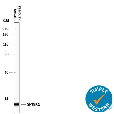
Simple Western lane view shows lysates of human pancreas tissue, loaded at 0.2 mg/mL. A specific band was detected for SPINK1 at approximately 5 kDa (as indicated) using 5 µg/mL of Sheep Anti-Human SPINK1 Antigen Affinity-purified Polyclonal Antibody (Catalog # AF7496) followed by 1:50 dilution of HRP-conjugated Anti-Sheep IgG Secondary Antibody (Catalog # HAF016). This experiment was conducted under reducing conditions and using the 12-230 kDa separation system.Detection of Human SPINK1 by Simple WesternTM.
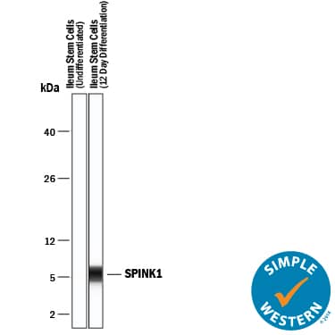
Simple Western lane view shows lysates of undifferentiated Ileum stem cells and day 12 differentiation Ileum stem cells, loaded at 0.2 mg/mL. A specific band was detected for SPINK1 at approximately 6 kDa (as indicated) using 5 µg/mL of Sheep Anti-Human SPINK1 Antigen Affinity-purified Polyclonal Antibody (Catalog # AF7496) followed by 1:50 dilution of HRP-conjugated Anti-Sheep IgG Secondary Antibody (Catalog # HAF016). This experiment was conducted under reducing conditions and using the 2-40 kDa separation system.Applications for Human SPINK1 Antibody
Simple Western
Sample: Human pancreas tissue and Day12 differentiation Ileum stem cells
Western Blot
Sample: Human pancreas tissue
Formulation, Preparation, and Storage
Purification
Reconstitution
Formulation
Shipping
Stability & Storage
- 12 months from date of receipt, -20 to -70 °C as supplied.
- 1 month, 2 to 8 °C under sterile conditions after reconstitution.
- 6 months, -20 to -70 °C under sterile conditions after reconstitution.
Background: SPINK1
SPINK1 (Serine Protease Inhibitor Kazal-type 1; also TATI and PST1) is a 6-7 kDa secreted polypeptide initially identified as a tumor-derived trypsin inhibitor. It is widely expressed, and found in cells diverse as pancreatic acinar cells, columnar cells of the stomach, renal collecting duct epithelium, and ureteric transitional plus breast epithelium. SPINK1 is known to be secreted with pancreatic zymogens, and apparently inactivates prematurely-activated trypsin, thus protecting the pancreas from trypsin-mediated enzyme activation. It also is reported to regulate cell migration and proliferation, the latter effect attributed to its structural resemblance to EGF, and its ability to bind to activate the EGFR. Mature human SPINK1 is 56 amino acids (aa) in length (aa 24-79). It contains one Kazal-like domain (aa 26-79) that possesses a potential proteolytic cleavage site between Lys41-Ile42. An 11-12 kDa form in SDS-PAGE has been reported for SPINK1, possibly reflecting dimerization. Mature human SPINK1 shares 66% aa sequence identity with mouse Spink3, the mouse equivalent to human SPINK1.
Long Name
Alternate Names
Gene Symbol
UniProt
Additional SPINK1 Products
Product Documents for Human SPINK1 Antibody
Product Specific Notices for Human SPINK1 Antibody
For research use only


