Human STING/TMEM173 Antibody
R&D Systems, part of Bio-Techne | Catalog # AF6516

Key Product Details
Species Reactivity
Validated:
Cited:
Applications
Validated:
Cited:
Label
Antibody Source
Product Specifications
Immunogen
Ala215-Ser379
Accession # Q86WV6
Specificity
Clonality
Host
Isotype
Scientific Data Images for Human STING/TMEM173 Antibody
Detection of Human STING/TMEM173 by Western Blot.
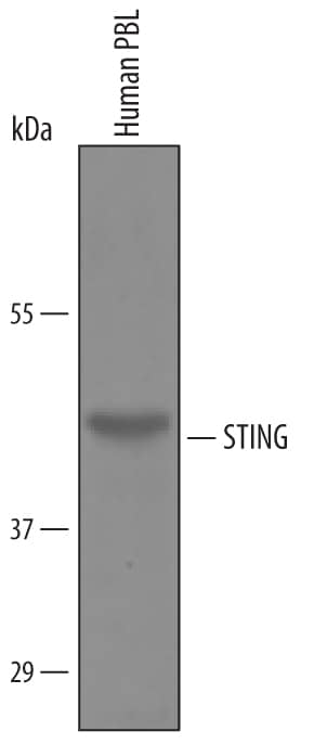
Western blot shows lysates of human peripheral blood lymphocytes (PBL). PVDF Membrane was probed with 1 µg/mL of Human/Mouse STING/TMEM173 Antigen Affinity-purified Polyclonal Antibody (Catalog # AF6516) followed by HRP-conjugated Anti-Sheep IgG Secondary Antibody (Catalog # HAF016). A specific band was detected for STING/TMEM173 at approximately 42 kDa (as indicated). This experiment was conducted under reducing conditions and using Immunoblot Buffer Group 8.Detection of Human STING/TMEM173 by Simple WesternTM.
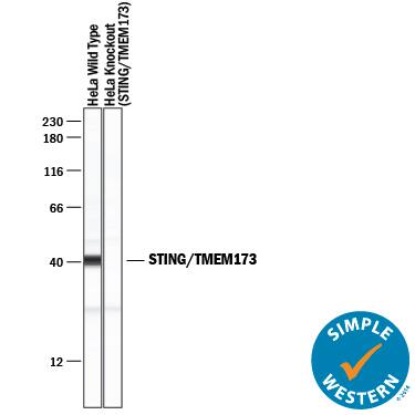
Simple Western lane view shows lysates of HeLa human cervical epithelial carcinoma parental cell line and STING/TMEM173 knockout HeLa cell line (KO), loaded at 0.2 mg/mL. A specific band was detected for STING/TMEM173 at approximately 41 kDa (as indicated) in the parental HeLa cell line, but is not detectable in knockout HeLa cell line. Sheep Anti-Human STING/TMEM173 Antigen Affinity-purified Polyclonal Antibody (Catalog # AF6516) was used at 10 µg/mL followed by 1:50 dilution of HRP-conjugated Anti-Sheep IgG Secondary Antibody (Catalog # HAF016). This experiment was conducted under reducing conditions and using the 12-230 kDa separation system.Detection of Mouse Human STING/TMEM173 Antibody by Immunohistochemistry
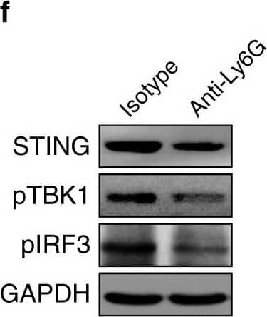
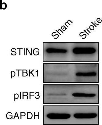
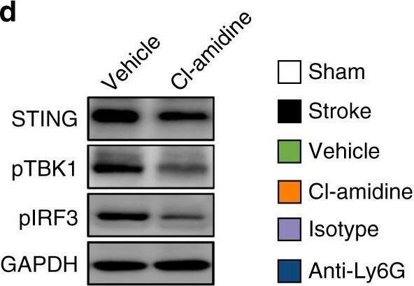
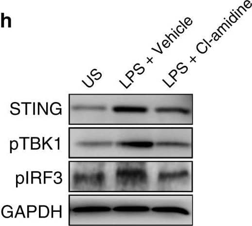
STING-mediated effects on vascular remodeling are due to NETs. i, j Confocal images of CD31-positive microvessels (i) and quantification of microvascular density (j) in the peri-infarct cortex at 14 days in mice treated with control IgG or IFNAR-neutralizing antibody, and STING shRNA or control adenovirus (n = 6), unpaired two-tailed Student’s t-test was applied with *P < 0.0001 (Vehicle vs. IFNAR), *P = 0.0007 (Ad-con vs. Ad-sh-STING). Bar = 40 μm. k, l In-vivo multiphoton microscopic images of perfused cortical capillaries with intravenously injected FITC-dextran (k) and quantification of perfused capillary length (l) at 14 days after stroke (n = 6), unpaired two-tailed Student’s t-test was applied with *P = 0.0006 (Vehicle vs. IFNAR), *P = 0.0080 (Ad-con vs. Ad-sh-STING). Bar = 100 μm. m, n In-vivo multiphoton microscopic images (m) of intravenously injected FITC-dextran leakage in cortical vessels at 14 days and quantification of the permeability (P) product of FITC-dextran (n) for each group (n = 6), unpaired two-tailed Student’s t-test was applied with *P = 0.0028 (Vehicle vs. IFNAR), *P = 0.0053 (Ad-con vs. Ad-sh-STING). Bar = 100 μm. Data are presented as mean ± SD. Source data underlying graph a–h, j, l, and n are provided as a Source Data file. Image collected and cropped by CiteAb from the following publication (https://pubmed.ncbi.nlm.nih.gov/32427863), licensed under a CC-BY license. Not internally tested by R&D Systems.Applications for Human STING/TMEM173 Antibody
Knockout Validated
Simple Western
Sample: HeLa human cervical epithelial carcinoma cell line
Western Blot
Sample: Human peripheral blood lymphocytes (PBL)
Formulation, Preparation, and Storage
Purification
Reconstitution
Formulation
Shipping
Stability & Storage
- 12 months from date of receipt, -20 to -70 °C as supplied.
- 1 month, 2 to 8 °C under sterile conditions after reconstitution.
- 6 months, -20 to -70 °C under sterile conditions after reconstitution.
Background: STING/TMEM173
STING (Stimulator of interferon genes; also ERIS, MPYS, MITA and TMEM173) is a 40-42 kDa 4-transmembrane protein that mediates both antiviral and MHC-II antigen recognition responses. STING is found in the ER, mitochondrial outer membrane and plasma membrane. It acts as an adaptor protein for intracellular viral detection molecules, participating in the induction of type I interferon. It also may play a role in the initiation of apoptosis following MHC-II engagement. Cells known to express STING include B cells, dendritic cells, macrophages, and monocytes. Human STING is 379 amino acids (aa) in length. It contains an N-terminal cytoplasmic region (aa 1-20), four transmembrane segments (aa 21-173), and a C-terminal cytoplasmic domain (aa 174-379). Ubiquitination occurs at Lys150, and phosphorylation occurs at Ser358. STING forms 80 kDa homodimers. There are two potential splice forms, one that shows a 25 aa substitution for aa 1-173, and another that possesses an alternative start site at Met215, coupled to a premature truncation following Arg334. Over aa 215-379, human STING shares 76% aa identity with mouse STING.
Long Name
Alternate Names
Gene Symbol
UniProt
Additional STING/TMEM173 Products
Product Documents for Human STING/TMEM173 Antibody
Product Specific Notices for Human STING/TMEM173 Antibody
For research use only
