Human TAFA1/FAM19A1 Antibody
R&D Systems, part of Bio-Techne | Catalog # AF5154

Key Product Details
Species Reactivity
Applications
Label
Antibody Source
Product Specifications
Immunogen
Ser26-Thr133
Accession # NP_998774
Specificity
Clonality
Host
Isotype
Scientific Data Images for Human TAFA1/FAM19A1 Antibody
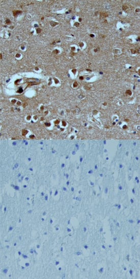
TAFA1/FAM19A1 in Human Brain.
TAFA1/FAM19A1 was detected in immersion fixed paraffin-embedded sections of human brain (cortex) using Goat Anti-Human TAFA1/FAM19A1 Antigen Affinity-purified Polyclonal Antibody (Catalog # AF5154) at 15 µg/mL overnight at 4 °C. Tissue was stained using the Anti-Goat HRP-DAB Cell & Tissue Staining Kit (brown; Catalog # CTS008) and counterstained with hematoxylin (blue). Lower panel shows a lack of labeling when primary antibodies are omitted and tissue is stained only with secondary antibody followed by incubation with detection reagents. View our protocol for Chromogenic IHC Staining of Paraffin-embedded Tissue Sections.Applications for Human TAFA1/FAM19A1 Antibody
Immunohistochemistry
Sample: Immersion fixed paraffin-embedded sections of human brain (cortex)
Western Blot
Sample: Recombinant Human TAFA1/FAM19A1 (Catalog # 5154-TA)
Neutralization
Formulation, Preparation, and Storage
Purification
Reconstitution
Formulation
Shipping
Stability & Storage
- 12 months from date of receipt, -20 to -70 °C as supplied.
- 1 month, 2 to 8 °C under sterile conditions after reconstitution.
- 6 months, -20 to -70 °C under sterile conditions after reconstitution.
Background: TAFA1/FAM19A1
TAFA1 (also FAM19A1) is a secreted, 13 kDa member of the FAM19/TAFA family of chemokine-like proteins (1). It is synthesized as a 133 amino acid (aa) precursor that contains a 19 aa signal sequence and a 114 aa mature chain. Like other members of the FAM19/TAFA family, mature TAFA1 contains 10 regularly spaced cysteine residues that follow the pattern CX7CCX13CXCX14CX11CX4CX5CX10C, in which C represents a conserved cysteine residue and X represents a noncysteine amino acid (1). Human TAFA1 is 100% aa identical to mouse TAFA1. TAFA1 is expressed exclusively in the brain, with highest expression in the frontal cortex, temporal cortex, occipital cortex, parietal cortex and medulla, and low levels in the basal ganglion, thalamus, and cerebellum (1). The biological functions of TAFA family members remain to be determined, but there are a few tentative hypotheses. First, TAFAs may modulate immune responses in the CNS by functioning as brain-specific chemokines, and may act with other chemokines to optimize the recruitment and activity of immune cells in the CNS (1). Second, TAFAs may represent a novel class of neurokines that act as regulators of immune nervous cells (1, 2). And third, TAFAs may control axonal sprouting following brain injury (1).
References
- Tang, Y.T. et al. (2004) Genomics 83:727.
- Benveniste, E. (1998) Cytokine Growth Factor Rev. 9:259.
Long Name
Alternate Names
Gene Symbol
UniProt
Additional TAFA1/FAM19A1 Products
Product Documents for Human TAFA1/FAM19A1 Antibody
Product Specific Notices for Human TAFA1/FAM19A1 Antibody
For research use only
