Human Thymidine Kinase 1 Antibody
R&D Systems, part of Bio-Techne | Catalog # MAB81801
Recombinant Monoclonal Antibody.

Key Product Details
Species Reactivity
Human
Applications
Simple Western, Western Blot
Label
Unconjugated
Antibody Source
Recombinant Monoclonal Rabbit IgG Clone # 2627A
Product Specifications
Immunogen
E. coli-derived human Thymidine Kinase 1
Ser2-Asn234
Accession # P04183
Ser2-Asn234
Accession # P04183
Specificity
Detects human Thymidine Kinase 1 in direct ELISAs.
Clonality
Monoclonal
Host
Rabbit
Isotype
IgG
Scientific Data Images for Human Thymidine Kinase 1 Antibody
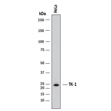
Detection of Human Thymidine Kinase 1 by Western Blot.
Western blot shows lysates of HeLa human cervical epithelial carcinoma cell line. PVDF membrane was probed with 1 µg/mL of Rabbit Anti-Human Thymidine Kinase 1 Monoclonal Antibody (Catalog # MAB81801) followed by HRP-conjugated Anti-Rabbit IgG Secondary Antibody (HAF008). A specific band was detected for Thymidine Kinase 1 at approximately 25 kDa (as indicated). This experiment was conducted under reducing conditions and using Western Blot Buffer Group 1.Detection of Human Thymidine Kinase 1 by Simple WesternTM.
Simple Western lane view shows lysates of A431 human epithelial carcinoma cell line, HeLa human cervical epithelial carcinoma cell line and SK-BR-3 human breast cancer cell line, loaded at 0.2 mg/mL. A specific band was detected for Thymidine Kinase 1 at approximately 32 kDa (as indicated) using 10 µg/mL of Rabbit Anti-Human Thymidine Kinase 1 Monoclonal Antibody (Catalog # MAB81801) . This experiment was conducted under reducing conditions and using the 12-230 kDa separation system.Applications for Human Thymidine Kinase 1 Antibody
Application
Recommended Usage
Simple Western
10 µg/mL
Sample: A431 human epithelial carcinoma cell line, HeLa human cervical epithelial carcinoma cell line and SK‑BR‑3 human breast cancer cell line
Sample: A431 human epithelial carcinoma cell line, HeLa human cervical epithelial carcinoma cell line and SK‑BR‑3 human breast cancer cell line
Western Blot
1 µg/mL
Sample: HeLa human cervical epithelial carcinoma cell line
Sample: HeLa human cervical epithelial carcinoma cell line
Formulation, Preparation, and Storage
Purification
Protein A or G purified from cell culture supernatant
Reconstitution
Reconstitute at 0.5 mg/mL in sterile PBS. For liquid material, refer to CoA for concentration.
Formulation
Lyophilized from a 0.2 μm filtered solution in PBS with Trehalose. *Small pack size (SP) is supplied either lyophilized or as a 0.2 µm filtered solution in PBS.
Shipping
Lyophilized product is shipped at ambient temperature. Liquid small pack size (-SP) is shipped with polar packs. Upon receipt, store immediately at the temperature recommended below.
Stability & Storage
Use a manual defrost freezer and avoid repeated freeze-thaw cycles.
- 12 months from date of receipt, -20 to -70 °C as supplied.
- 1 month, 2 to 8 °C under sterile conditions after reconstitution.
- 6 months, -20 to -70 °C under sterile conditions after reconstitution.
Background: Thymidine Kinase 1
References
- Wintersberger, E. (1997) Biochem. Soc. Trans. 25:303.
- Littelfield, J.W. (1966) Biochim. Biophys. Acta 114:398.
- Flemington, E, et al. (1987) Gene 52:267.
- Berk, A.J. et al. (1973) Arch. Biochem. Biophys. 154:563.
- Zhu, C. et al. (2006) Nucleosides Nucleotides Nucleic Acids 25:1185.
- Munch-Petersen, B. et al. (1993) J. Biol. Chem. 268:15621.
- Mikkelsen, N. E. et al. (2003). Biochemistry 42:5706.
- Doi S, et al. (1990) Nagoya J. Med. Sci. 52:19.
- Ellims PH, et al. (1981). Cancer Res. 41: 691.
- Lin TS, et al. (1976) J. Med. Chem.19:495.
- Baba M, et al. (1987). Biochem. Biophys. Res. Commun. 142:128.
- Wu, Z.L. (2011) PLoS One 6:e23172.
Alternate Names
TK1
Gene Symbol
TK1
UniProt
Additional Thymidine Kinase 1 Products
Product Documents for Human Thymidine Kinase 1 Antibody
Product Specific Notices for Human Thymidine Kinase 1 Antibody
For research use only
Loading...
Loading...
Loading...
Loading...

