Human TMEM119 Antibody
R&D Systems, part of Bio-Techne | Catalog # MAB103131


Key Product Details
Species Reactivity
Applications
Label
Antibody Source
Product Specifications
Immunogen
Arg26-Met96
Accession # Q4V9L6.1
Specificity
Clonality
Host
Isotype
Scientific Data Images for Human TMEM119 Antibody
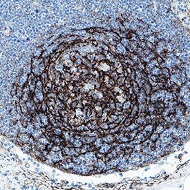
TMEM119 in Human lymph node.
TMEM119 was detected in immersion fixed paraffin-embedded sections of human lymph node using Rabbit Anti-Human TMEM119 Monoclonal Antibody (Catalog # MAB103131) at 1 µg/mL for 1 hour at room temperature followed by incubation with the Anti-Rabbit IgG VisUCyte™ HRP Polymer Antibody (VC003). Before incubation with the primary antibody, tissue was subjected to heat-induced epitope retrieval using Antigen Retrieval Reagent-Basic (CTS013). Tissue was stained using DAB (brown) and counterstained with hematoxylin (blue). Specific staining was localized to cell membrane and cytoplasm in lymphocytes. Staining was performed using our protocol for IHC Staining with VisUCyte HRP Polymer Detection Reagents.Detection of TMEM119 in HEK293 Human Cell Line Transfected with Human TMEM119 and eGFP by Flow Cytometry
HEK293 human embryonic kidney cell line transfected with (A) human TMEM119 or (B) irrelevant protein, and eGFP was stained with Rabbit Anti-Human TMEM119 Monoclonal Antibody (Catalog # MAB103131) followed by Allophycocyanin-conjugated Anti-Rabbit IgG Secondary Antibody (F0111). Quadrant markers were set based on Rabbit IgG Isotype Control (MAB1050, data not shown). Staining was performed using our Staining Membrane-associated Proteins protocol.Applications for Human TMEM119 Antibody
Flow Cytometry
Sample: HEK293 Human Cell Line Transfected with Human TMEM119 and eGFP
Immunohistochemistry
Sample: Immersion fixed paraffin-embedded sections of human lymph node
Formulation, Preparation, and Storage
Purification
Reconstitution
Formulation
Shipping
Stability & Storage
- 12 months from date of receipt, -20 to -70 °C as supplied.
- 1 month, 2 to 8 °C under sterile conditions after reconstitution.
- 6 months, -20 to -70 °C under sterile conditions after reconstitution.
Background: TMEM119
TMEM119 (Transmembrane Protein 119, also known as Osteoblast Induction Factor or OBIF), is an approximately 38-kDa type 1 transmembrane protein that is predominantly expressed in osteoblasts and is upregulated during osteoblastic differentiation (1, 2). TMEM119 is also expressed in a cell line of microglia, and TMEM119 immunoreactivity is observed in a specific subset of microglia in brains of neurodegenerative diseases, such as Alzheimers disease (3). Mature human TMEM119 consists of a 71 amino acid (aa) extracellular domain (ECD), a 21 aa transmembrane segment, and a 166 aa cytoplasmic domain. Within the ECD, human TMEM119 shares 78% and 75% aa sequence identity with mouse and rat TMEM119, respectively. TMEM-119 is involved in the osteoblast differentiation and bone development by acting as a ligand and has been reported to contribute to the proliferation, migration, and invasion of osteosarcoma cells, as well as functioning as an oncogene in osteosarcoma (3, 4).
References
- Jiang, Z,H. et al. (2017) Expt & Mol Med. 49:e329.
- Mizuhashi, K. et al. (2012) Dev. Growth Differ. 54:474.
- Satoh, J. et al. (2016) Neuropathol. 36:39.
- Kanamoto, T. et al. (2009) BMC Develop. Biol. 9:70.
Long Name
Alternate Names
Gene Symbol
UniProt
Additional TMEM119 Products
Product Documents for Human TMEM119 Antibody
Product Specific Notices for Human TMEM119 Antibody
For research use only
