Human VCAM-1/CD106 Antibody
R&D Systems, part of Bio-Techne | Catalog # AF809


Key Product Details
Validated by
Species Reactivity
Validated:
Cited:
Applications
Validated:
Cited:
Label
Antibody Source
Product Specifications
Immunogen
Extracellular domain
Specificity
Clonality
Host
Isotype
Endotoxin Level
Scientific Data Images for Human VCAM-1/CD106 Antibody
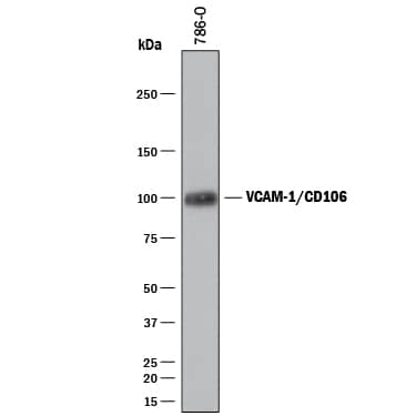
Detection of Human VCAM‑1/CD106 by Western Blot.
Western blot shows lysates of 786-O human renal cell adenocarcinoma cell line. PVDF membrane was probed with 2 µg/mL of Sheep Anti-Human VCAM-1/ CD106 Antigen Affinity-purified Polyclonal Antibody (Catalog # AF809) followed by HRP-conjugated Anti-Sheep IgG Secondary Antibody (Catalog # HAF016). A specific band was detected for VCAM-1/CD106 at approximately 100 kDa (as indicated). This experiment was conducted under reducing conditions and using Immunoblot Buffer Group 1.Detection of Human VCAM-1/CD106 by Simple WesternTM.
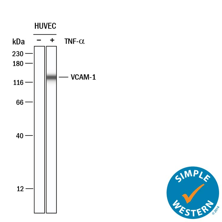
Simple Western lane view shows lysates of HUVEC human umbilical vein endothelial cells untreated (-) or treated (+) with 10 ng/ml Recombinant Human TNF-alpha (210-TA) for 24 hrs, loaded at 0.2 mg/mL. A specific band was detected for VCAM-1/CD106 at approximately 132 kDa (as indicated) using 20 µg/mL of Sheep Anti-Human VCAM-1/CD106 Antigen Affinity-purified Polyclonal Antibody (Catalog # AF809) followed by 1:50 dilution of HRP-conjugated Anti-Sheep IgG Secondary Antibody (HAF016). This experiment was conducted under reducing conditions and using the 12-230 kDa separation system.Applications for Human VCAM-1/CD106 Antibody
Adhesion Blockade
Simple Western
Sample: HUVEC human umbilical vein endothelial cells
Western Blot
Sample: 786‑O human renal cell adenocarcinoma cell line
Reviewed Applications
Read 3 reviews rated 4.3 using AF809 in the following applications:
Formulation, Preparation, and Storage
Purification
Reconstitution
Formulation
Shipping
Stability & Storage
- 12 months from date of receipt, -20 to -70 °C as supplied.
- 1 month, 2 to 8 °C under sterile conditions after reconstitution.
- 6 months, -20 to -70 °C under sterile conditions after reconstitution.
Background: VCAM-1/CD106
VCAM-1 (CD106), a member of the immunoglobulin superfamily, is a cell surface protein expressed by activated endothelial cells and certain leukocytes (such as macrophages). VCAM-1 expression is induced by IL-1 beta, IL-4, TNF-alpha, and IFN-gamma. VCAM-1 binds to leukocyte integrins VLA-4 and alpha4 beta7. The human and mouse VCAM-1 proteins share approximately 76% amino acid similarity.
During the inflammatory adhesion mechanism, activated integrins halt rolling leukocytes and attach them firmly to the vascular endothelium. They do this by binding to their ligands, for example VCAM-1, on endothelium. The VCAM-1: VLA-4/ alpha4 beta7 interaction is also thought to be involved in the extravasation of white blood cells through the blood vessel wall to sites of inflammation.
ELISA techniques have shown that detectable levels of soluble VCAM-1 are present in the biological fluids of apparently normal individuals. Furthermore, a number of studies have reported that levels of VCAM-1 may be elevated or lowered in subjects with a variety of pathological conditions.
Long Name
Alternate Names
Gene Symbol
Additional VCAM-1/CD106 Products
Product Documents for Human VCAM-1/CD106 Antibody
Product Specific Notices for Human VCAM-1/CD106 Antibody
For research use only
