Human VEGF165b Antibody
R&D Systems, part of Bio-Techne | Catalog # MAB3045

Key Product Details
Species Reactivity
Validated:
Human
Cited:
Human, Mouse, Bovine
Applications
Validated:
Immunofluorescence, Immunohistochemistry, Western Blot
Cited:
ELISA Development, Flow Cytometry, Immunohistochemistry, Immunohistochemistry-Paraffin, Neutralization, Western Blot
Label
Unconjugated
Antibody Source
Monoclonal Mouse IgG1 Clone # 56-1
Product Specifications
Immunogen
KLH-conjugated human VEGF165b synthetic peptide
TCRSLTRKD
Accession # AAL27435
TCRSLTRKD
Accession # AAL27435
Specificity
Detects human VEGF165b in direct ELISAs and Western blots. Recognizes an epitope within a 9 amino acid sequence at the C-terminus of human VEGF165b. In direct ELISAs and Western blots, no cross-reactivity with recombinant human (rh) VEGF206, rhVEGF-B167, rhVEGF-B186, rhVEGF-C, or rhVEGF-D is observed.
Clonality
Monoclonal
Host
Mouse
Isotype
IgG1
Scientific Data Images for Human VEGF165b Antibody
VEGF165bin Human Placenta.
VEGF165bwas detected in immersion fixed paraffin-embedded sections of human placenta using Mouse Anti-Human VEGF165bMonoclonal Antibody (Catalog # MAB3045) at 15 µg/mL overnight at 4 °C. Tissue was stained using the Anti-Mouse HRP-DAB Cell & Tissue Staining Kit (brown; Catalog # CTS002) and counterstained with hematoxylin (blue). Specific staining was localized to syncytiotrophoblasts. View our protocol for Chromogenic IHC Staining of Paraffin-embedded Tissue Sections.VEGF expression determined by Western blot
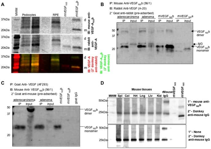
VEGF expression determined by Western blot and immunoprecipitation.A. Western blot using LiCor Odyssey to simultaneously image pan-VEGF and VEGF-A165b probed western blot. Two different podocyte samples, and a primary RPE sample were run on a gel and probed with antibodies to VEGF-A165b (mouse monoclonal anti-CTRSLTRKD, and 680nm-donkey anti-mouse, top image) and pan-VEGF (rabbit polyclonal anti-VEGF, and 800nm-donkey anti-rabbit, middle image). The bottom image is the pseudocoloured combined image (600nm green, 800nm red). Note the red VEGF165, but yellow VEGF-A165b. MWM = molecular weight marker. d = dimer, m = monomer. B. Protein extracted from human cell lines (adenoma and adenocarcinoma(AC)) subjected to immunoprecipitation (IP) for VEGF-A165b and immunoblotting (IB) for total VEGF-A. A clear strong band was seen in the IP for both cell types at ∼23kDa and ∼46kDa, consistent with the IP for recombinant human VEGF-A165b. A weaker band was seen in the input protein (not subjected to IP), and a second band slightly higher in the AC. A weak band at approximately 56kDa and 28kDa was seen in all lanes subjected to IP, including the VEGF-A165a band, but not seen in the recombinant human VEGF-A165b not subjected to IP, indicating that this is cross reactivity with the IgG. This band was clearly above the VEGF-A165b bands. C. Protein extracted from human cell lines (adenoma and adenocarcinoma(AC)) subjected to immunoprecipitation (IP) for VEGF-A and immunoblotting (IB) for VEGF-A165b. A clear strong band was seen in the IP for both cell types at ∼23kDa, the same size as recombinant human VEGF-A165b. In the input a band at ∼46Da was seen predominantly, for both cell types, labelled as VEGF-A165b dimers. D. Mouse tissues probed with VEGF-A165b antibody detect mouse IgG due to the secondary antibody. Top image, western blot of mouse tissues, recombinant mouse IgG or human VEGF-A165b or VEGF-A165b probed with mouse anti-CTRSLTRKD, and 680nm-donkey anti-mouse IgG. Bottom image blot of same tissues, probed without primary antibody. The same bands are seen in the mouse tissues. Spl = spleen, Col = colon, Hrt = heart, Lng = lung, Liv = liver, Kid = kidney. Image collected and cropped by CiteAb from the following publication (https://pubmed.ncbi.nlm.nih.gov/23935865), licensed under a CC-BY license. Not internally tested by R&D Systems.Applications for Human VEGF165b Antibody
Application
Recommended Usage
Immunofluorescence
Woolard, J. et al. (2004) Canc. Res. 64:7822.
Immunohistochemistry
Bates, D.O. et al. (2006) Clinical Sci. 110:575.
Western Blot
1 µg/mL
Sample: Recombinant Human VEGF165b (Catalog # 3045-VE)
Sample: Recombinant Human VEGF165b (Catalog # 3045-VE)
Reviewed Applications
Read 2 reviews rated 5 using MAB3045 in the following applications:
Formulation, Preparation, and Storage
Purification
Protein A or G purified from hybridoma culture supernatant
Reconstitution
Reconstitute at 0.5 mg/mL in sterile PBS. For liquid material, refer to CoA for concentration.
Formulation
Lyophilized from a 0.2 μm filtered solution in PBS with Trehalose. *Small pack size (SP) is supplied either lyophilized or as a 0.2 µm filtered solution in PBS.
Shipping
Lyophilized product is shipped at ambient temperature. Liquid small pack size (-SP) is shipped with polar packs. Upon receipt, store immediately at the temperature recommended below.
Stability & Storage
Use a manual defrost freezer and avoid repeated freeze-thaw cycles.
- 12 months from date of receipt, -20 to -70 °C as supplied.
- 1 month, 2 to 8 °C under sterile conditions after reconstitution.
- 6 months, -20 to -70 °C under sterile conditions after reconstitution.
Background: VEGF
Long Name
Vascular Endothelial Growth Factor
Alternate Names
MVCD1, VAS, Vasculotropin, VEGF-A, VEGFA, VPF
Entrez Gene IDs
Gene Symbol
VEGFA
UniProt
Additional VEGF Products
Product Documents for Human VEGF165b Antibody
Product Specific Notices for Human VEGF165b Antibody
For research use only
Loading...
Loading...
Loading...
Loading...

