Human VEGFR2/KDR/Flk‑1 Biotinylated Antibody Best Seller
R&D Systems, part of Bio-Techne | Catalog # BAF357


Key Product Details
Species Reactivity
Validated:
Cited:
Applications
Validated:
Cited:
Label
Antibody Source
Product Specifications
Immunogen
Ala20-Glu764
Accession # AAC16450
Specificity
Clonality
Host
Isotype
Scientific Data Images for Human VEGFR2/KDR/Flk‑1 Biotinylated Antibody
Detection of Human VEGFR2/KDR/Flk-1 by ELISA
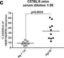
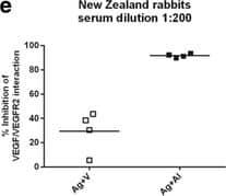
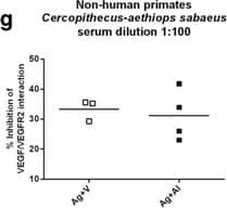
Humoral response in pre-clinical models. Specific IgG antibodies were detected by ELISA using GST-hVEGF as coating antigen (a, b, d and f). The ability of animal sera antibodies to block VEGF/VEGFR2 interaction was determined using a competitive ELISA, where a soluble VEGFR2 competes with diluted serum in plates coated with GST-hVEGF (c, e and g). CIGB-247 combinations using VSSP or aluminum phosphate as adjuvants were administered weekly (eight vaccinations) or bi-weekly (four vaccinations) respectively. Horizontal bars represent the mean values of antibody titer or blocking activity, which are shown for each group. p-Values were calculated according to unpaired t-test. Reference dose (A + V): 400 μg of antigen + 200 μg of VSSP; (¼Ag + ½ V): 100 μg of antigen + 100 μg of VSSP; (¼Ag + V): 100 μg of antigen + 200 μg of VSSP; (Ag + Al): 400 μg of antigen + aluminum phosphate Image collected and cropped by CiteAb from the following publication (https://pubmed.ncbi.nlm.nih.gov/28747172), licensed under a CC-BY license. Not internally tested by R&D Systems.Detection of Human VEGFR2/KDR/Flk-1 by ELISA
Humoral response in pre-clinical models. Specific IgG antibodies were detected by ELISA using GST-hVEGF as coating antigen (a, b, d and f). The ability of animal sera antibodies to block VEGF/VEGFR2 interaction was determined using a competitive ELISA, where a soluble VEGFR2 competes with diluted serum in plates coated with GST-hVEGF (c, e and g). CIGB-247 combinations using VSSP or aluminum phosphate as adjuvants were administered weekly (eight vaccinations) or bi-weekly (four vaccinations) respectively. Horizontal bars represent the mean values of antibody titer or blocking activity, which are shown for each group. p-Values were calculated according to unpaired t-test. Reference dose (A + V): 400 μg of antigen + 200 μg of VSSP; (¼Ag + ½ V): 100 μg of antigen + 100 μg of VSSP; (¼Ag + V): 100 μg of antigen + 200 μg of VSSP; (Ag + Al): 400 μg of antigen + aluminum phosphate Image collected and cropped by CiteAb from the following publication (https://pubmed.ncbi.nlm.nih.gov/28747172), licensed under a CC-BY license. Not internally tested by R&D Systems.Detection of Human VEGFR2/KDR/Flk-1 by ELISA
Humoral response in pre-clinical models. Specific IgG antibodies were detected by ELISA using GST-hVEGF as coating antigen (a, b, d and f). The ability of animal sera antibodies to block VEGF/VEGFR2 interaction was determined using a competitive ELISA, where a soluble VEGFR2 competes with diluted serum in plates coated with GST-hVEGF (c, e and g). CIGB-247 combinations using VSSP or aluminum phosphate as adjuvants were administered weekly (eight vaccinations) or bi-weekly (four vaccinations) respectively. Horizontal bars represent the mean values of antibody titer or blocking activity, which are shown for each group. p-Values were calculated according to unpaired t-test. Reference dose (A + V): 400 μg of antigen + 200 μg of VSSP; (¼Ag + ½ V): 100 μg of antigen + 100 μg of VSSP; (¼Ag + V): 100 μg of antigen + 200 μg of VSSP; (Ag + Al): 400 μg of antigen + aluminum phosphate Image collected and cropped by CiteAb from the following publication (https://pubmed.ncbi.nlm.nih.gov/28747172), licensed under a CC-BY license. Not internally tested by R&D Systems.Applications for Human VEGFR2/KDR/Flk‑1 Biotinylated Antibody
Western Blot
Sample: Recombinant Human VEGFR2/KDR/Flk‑1 Fc Chimera (Catalog # 357-KD)
Human VEGFR2/KDR Sandwich Immunoassay
Formulation, Preparation, and Storage
Purification
Reconstitution
Formulation
Shipping
Stability & Storage
- 12 months from date of receipt, -20 to -70 °C as supplied.
- 1 month, 2 to 8 °C under sterile conditions after reconstitution.
- 6 months, -20 to -70 °C under sterile conditions after reconstitution.
Background: VEGFR2/KDR/Flk-1
VEGFR2 (KDR/Flk-1), VEGFR1 (Flt-1) and VEGFR3 (Flt-4) belong to the class III subfamily of receptor tyrosine kinases (RTKs). All three receptors contain seven immunoglobulin-like repeats in their extracellular domains and kinase insert domains in their intracellular regions. The expression of VEGFR1, 2, and 3 is almost exclusively restricted to the endothelial cells. These receptors are likely to play essential roles in vasculogenesis and angiogenesis.
VEGFR2 cDNA encodes a 1356 amino acid (aa) residue precursor protein with a 19 aa residue signal peptide. Mature VEGFR2 is composed of a 745 aa residue extracellular domain, a 25 aa residue transmembrane domain and a 567 aa residue cytoplasmic domain. In contrast to VEGFR1 which binds both PlGF and VEGF with high affinity, VEGFR2 binds VEGF but not PlGF with high affinity.
References
- Ferra, N. and R. Davis-Smyth (1997) Endocrine Reviews 18:4.
Long Name
Alternate Names
Gene Symbol
UniProt
Additional VEGFR2/KDR/Flk-1 Products
Product Documents for Human VEGFR2/KDR/Flk‑1 Biotinylated Antibody
Product Specific Notices for Human VEGFR2/KDR/Flk‑1 Biotinylated Antibody
For research use only

