Human ZAG Antibody
R&D Systems, part of Bio-Techne | Catalog # AF4764


Key Product Details
Species Reactivity
Applications
Label
Antibody Source
Product Specifications
Immunogen
Gln21-Ser298
Accession # Q5XKQ4
Specificity
Clonality
Host
Isotype
Scientific Data Images for Human ZAG Antibody
Detection of Human ZAG by Western Blot.
Western blot shows lysates of human prostate tissue. PVDF membrane was probed with 1 µg/mL of Goat Anti-Human ZAG Antigen Affinity-purified Polyclonal Antibody (Catalog # AF4764) followed by HRP-conjugated Anti-Goat IgG Secondary Antibody (Catalog # HAF019). A specific band was detected for ZAG at approximately 40 kDa (as indicated). This experiment was conducted under reducing conditions and using Immunoblot Buffer Group 8.ZAG in Human Breast Cancer Tissue.
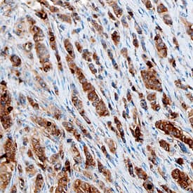
ZAG was detected in immersion fixed paraffin-embedded sections of human breast cancer tissue using Goat Anti-Human ZAG Antigen Affinity-purified Polyclonal Antibody (Catalog # AF4764) at 10 µg/mL overnight at 4 °C. Before incubation with the primary antibody, tissue was subjected to heat-induced epitope retrieval using Antigen Retrieval Reagent-Basic (Catalog # CTS013). Tissue was stained using the Anti-Goat HRP-DAB Cell & Tissue Staining Kit (brown; Catalog # CTS008) and counterstained with hematoxylin (blue). Specific staining was localized to epithelial cells. View our protocol for Chromogenic IHC Staining of Paraffin-embedded Tissue Sections.ZAG in Human Mesenchymal Stem Cells.
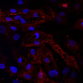
ZAG was detected in immersion fixed human mesenchymal stem cells differentiated into adipocytes using Goat Anti-Human ZAG Antigen Affinity-purified Polyclonal Antibody (Catalog # AF4764) at 10 µg/mL for 3 hours at room temperature. Cells were differentiated using using StemXVivo Osteogenic/Adipogenic Base Media (Catalog # CCM007) and StemXVivo Adipogenic Supplement (100X) (Catalog # CCM011). Cells were stained using the NorthernLights™ 557-conjugated Anti-Goat IgG Secondary Antibody (red; Catalog # NL001) and counterstained with DAPI (blue). Specific staining was localized to cytoplasm. View our protocol for Fluorescent ICC Staining of Stem Cells on Coverslips.Applications for Human ZAG Antibody
Immunocytochemistry
Sample: Immersion fixed human mesenchymal stem cells differentiated into adipocytes using StemXVivo Osteogenic/Adipogenic Base Media (Catalog # CCM007) and StemXVivo Adipogenic Supplement (100X) (Catalog # CCM011)
Immunohistochemistry
Sample: Immersion fixed paraffin-embedded sections of human breast cancer tissue
Western Blot
Sample: Human prostate tissue
Formulation, Preparation, and Storage
Purification
Reconstitution
Formulation
Shipping
Stability & Storage
- 12 months from date of receipt, -20 to -70 °C as supplied.
- 1 month, 2 to 8 °C under sterile conditions after reconstitution.
- 6 months, -20 to -70 °C under sterile conditions after reconstitution.
Background: ZAG
ZAG (zinc-alpha 2-glycoprotein; also ZA2G) is a 40 kDa, secreted member of the MHC class I family of proteins. It is produced by adipocytes and various epithelial cells that generate exocrine-type secretions. ZAG is reported to stimulate lipid breakdown and thus may play a role in lipid homeostasis. Mature human ZAG is 278 amino acids (aa) in length. It contains one MHC class I antigen region (aa 26‑201) and a C2‑type Ig‑like domain (aa 207‑292). Two alternate splice forms exist; one shows a 66 aa substitution for the C‑terminal 30 aa, and a second shows a nine Lys substitution for aa 151‑298. Mature human ZAG is 60% aa identical to mouse ZAG.
Long Name
Alternate Names
Gene Symbol
UniProt
Additional ZAG Products
Product Documents for Human ZAG Antibody
Product Specific Notices for Human ZAG Antibody
For research use only

