LAG-3 Antibody (L4-PL33) - BSA Free
Novus Biologicals, part of Bio-Techne | Catalog # NBP3-23442


Conjugate
Catalog #
Key Product Details
Species Reactivity
Human, Primate
Applications
ELISA, Flow Cytometry, Immunocytochemistry/ Immunofluorescence, Immunohistochemistry, Immunohistochemistry-Frozen, Immunohistochemistry-Paraffin, Immunoprecipitation, Western Blot
Label
Unconjugated
Antibody Source
Monoclonal Mouse IgG1 kappa Clone # L4-PL33
Format
BSA Free
Concentration
1 mg/ml
Product Specifications
Immunogen
This LAG-3 Antibody (L4-PL33) was prepared from synthetic peptide corresponding to 30 aa (GPPAAAPGHPLAPGPHPAAPSSWGPRPRRY) from the first N-terminal D1 domain of human LAG-3 (lymphocyte activation gene-3).
Reactivity Notes
Primate reactivity reported in scientific literature (PMID: 32284611).
Clonality
Monoclonal
Host
Mouse
Isotype
IgG1 kappa
Scientific Data Images for LAG-3 Antibody (L4-PL33) - BSA Free
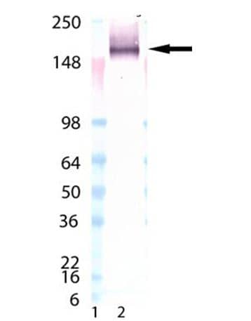
Western Blot-NBP3-23442-
Western Blot-NBP3-23442-Analysis of LAG-3 mAb (L4-PL33) : Lane 1: MW marker, Lane 2: LAG-3 (human):Fc (human), (recombinant)IHC-P-NBP3-23442-
IHC-P-NBP3-23442-Analysis of formalin-fixed, paraffin-embedded human tonsil tissue stained with LAG-3 (human) recombinant mAb (L4-PL33) at 6ug/ml.Flow Cytometry-NBP3-23442-
Flow Cytometry-NBP3-23442-Analysis of 0.5x10^6 THP-1 cells stained using LAG-3 mAb (L4-PL33) at a concentration of 5μg/ml, followed by Alexa Fluor® 488 conjugated anti-mouse secondary antibody.Applications for LAG-3 Antibody (L4-PL33) - BSA Free
Application
Recommended Usage
Immunocytochemistry/ Immunofluorescence
1:10 - 1:500
Immunohistochemistry
1:10 - 1:500
Immunohistochemistry-Frozen
1:150
Immunoprecipitation
10 ug/mL
Western Blot
5 ug/mL
Formulation, Preparation, and Storage
Purification
Protein A or G purified
Formulation
PBS, 10% glycerol
Format
BSA Free
Preservative
0.02% Sodium Azide
Concentration
1 mg/ml
Shipping
The product is shipped with polar packs. Upon receipt, store it immediately at the temperature recommended below.
Stability & Storage
Store at 4C short term. Aliquot and store at -20C long term. Avoid freeze-thaw cycles.
Background: LAG-3
As mentioned above, LAG-3 binds to MHCII and this occurs via a proline-rich amino acid loop in D1 (1, 3). Another unique feature of LAG-3 is the longer connecting peptide region between the D4 and the transmembrane, which is acted upon and cleaved by metalloproteinases a disintegrin and metallopeptidase domain (ADAM) 10 and ADAM17 to generate a soluble 54 kDa form of LAG-3 (sLAG-3) (1, 3). The interaction of LAG-3 with MHCII prevents the MHC molecule from binding to a T-cell receptor (TCR) or CD4, thereby functioning in an inhibitory role and suppressing the TCR signal (4). When LAG-3 crosslinks with the TCR/CD3 complex, it causes reduced T-cell proliferation and cytokine secretion (4). This negative regulation is important in controlling autoimmunity as one study found Lag3-/- NOD (non-obese diabetic) mice had accelerated diabetes onset and increased T-cell infiltration into islet cells (5). On the other hand, besides being a negative regulator of T-cells, LAG-3 binding to MHCII molecules on APCs induces dendritic cell maturation and cytokine secretion by monocytes (5, 6). In addition to MHCII, other reported ligands for LAG-3 includes fibrinogen-like protein 1 (FGL1), liver endothelial cell lectin (lSECtin), galectin-3 (Gal-3), and alpha-synuclein fibrils (1). Gal-3, for instance, is expressed on stromal cells and CD8+ T-cells in the tumor microenvironment and the interaction with LAG-3 was shown to be crucial for the suppression of secreted cytokine IFN-gamma and may control anti-tumor immune responses (1, 5). Interestingly, a mouse model of Parkinson's disease revealed LAG-3 binding to alpha-synuclein fibrils in the central nervous system, contributing to its pathogenesis (1, 5).
Recent cancer immunotherapeutic approaches have focused on inhibitory receptors such as LAG-3 to revive expression of cytotoxic T-cells to attack tumors (6). LAG-3 has been shown to be co-expressed and have synergy with another immune-checkpoint molecule called programmed-death 1 (PD-1) (1, 4, 5, 6). In a mouse model of colon adenocarcinoma LAG3 blockade alone was largely ineffective, however co-blockade of LAG-3 and PD-1 limited tumor growth and resulted in tumor clearance in 80% of mice, compared to 40% with PD-1 blockade alone (5). Additionally, in a model of fibrosarcoma the LAG-3/PD-1 duel blockade increased survival and the percentage of tumor-free mice (5). Analysis of a variety of human tumor samples (e.g. melanoma, colon cancer, head and neck squamous cell carcinoma) also suggest that LAG3 alone and combinatorial treatment with PD-1 may be a good target for treatment (1, 3-6). To date there are over 10 different agents targeting LAG-3 in clinical trials for cancer either as an anti-LAG-3 blocking antibody monotherapy or as a combination antagonist bispecific antibody, primarily with PD-1 (1, 3-6).
Alternative names for LAG-3 includes 17b4 lag3, 17b4 neutralizing, 17b4, CD223, FDC, LAG-3 17b4, LAG-3 blocking, and LAG3.
References
1. Maruhashi, T., Sugiura, D., Okazaki, I. M., & Okazaki, T. (2020). LAG-3: from molecular functions to clinical applications. Journal for Immunotherapy of Cancer, 8(2), e001014. https://doi.org/10.1136/jitc-2020-001014
2. Triebel, F., Jitsukawa, S., Baixeras, E., Roman-Roman, S., Genevee, C., Viegas-Pequignot, E., & Hercend, T. (1990). LAG-3, a novel lymphocyte activation gene closely related to CD4. The Journal of experimental medicine, 171(5), 1393-1405. https://doi.org/10.1084/jem.171.5.1393
3. Ruffo, E., Wu, R. C., Bruno, T. C., Workman, C. J., & Vignali, D. (2019). Lymphocyte-activation gene 3 (LAG3): The next immune checkpoint receptor. Seminars in immunology, 42, 101305. https://doi.org/10.1016/j.smim.2019.101305
4. Long, L., Zhang, X., Chen, F., Pan, Q., Phiphatwatchara, P., Zeng, Y., & Chen, H. (2018). The promising immune checkpoint LAG-3: from tumor microenvironment to cancer immunotherapy. Genes & cancer, 9(5-6), 176-189.
5. Andrews, L. P., Marciscano, A. E., Drake, C. G., & Vignali, D. A. (2017). LAG3 (CD223) as a cancer immunotherapy target. Immunological reviews, 276(1), 80-96. https://doi.org/10.1111/imr.12519
6. Goldberg, M. V., & Drake, C. G. (2011). LAG-3 in Cancer Immunotherapy. Current topics in microbiology and immunology, 344, 269-278. https://doi.org/10.1007/82_2010_114
Long Name
Lymphocyte-activation Gene 3
Alternate Names
CD223, LAG3, 17b4, 17b4 lag3, 17b4 neutralizing, anti human lag3, Anti-LAG-3 Antibody, CD223 Antibody, CD223 Antigen Antibody, Human LAG3 Antibody, Human LAG-3 Antibody, LAG-3 17b4, LAG-3 blocking
Gene Symbol
LAG3
Additional LAG-3 Products
Product Documents for LAG-3 Antibody (L4-PL33) - BSA Free
Product Specific Notices for LAG-3 Antibody (L4-PL33) - BSA Free
This product is for research use only and is not approved for use in humans or in clinical diagnosis. Primary Antibodies are guaranteed for 1 year from date of receipt.
Loading...
Loading...
Loading...
Loading...
Loading...

