Laminin Antibody [Alexa Fluor® 647]
Novus Biologicals, part of Bio-Techne | Catalog # NB300-144AF647


Key Product Details
Species Reactivity
Validated:
Cited:
Applications
Validated:
Cited:
Label
Antibody Source
Concentration
Product Specifications
Immunogen
Reactivity Notes
Localization
Specificity
Marker
Clonality
Host
Isotype
Scientific Data Images for Laminin Antibody [Alexa Fluor® 647]
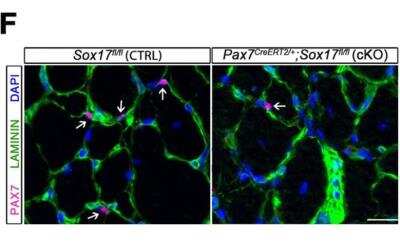
Frozen Mouse Tissue Stained for Laminin
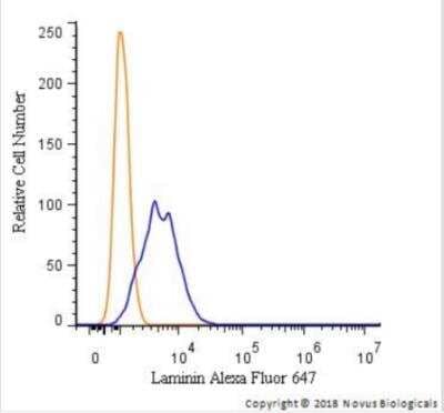
SOX17 regulates adult muscle regeneration after injury in Pax7CreERT2/+;Sox17fl/fl mutant mice. Representative images of cryosections from regenerating adult TA muscles d28 after injury, showing immunofluorescence for PAX7+ (quiescent, arrows) cells. Scale bar, 25 Aum. Image collected and cropped by Citeab from the following publication (SOXF factors regulate murine satellite cell self-renewal and function through inhibition ofI2-catenin activity. Elife (2018)) licensed under a CC-BY license.Flow Cytometry of HeLa Cells Stained with Conjugated Laminin Antibody
A surface stain was performed on HeLa cells with Laminin Antibody NB300-144AF647 (blue) and a matched isotype control (orange). Cells were incubated in an antibody dilution of 2.5 ug/mL for 20 minutes at room temperature. Both antibodies were conjugated to Alexa Fluor 647.Immunocytochemistry/ Immunofluorescence: Laminin Antibody [Alexa Fluor® 647] [NB300-144AF647] -
Immunocytochemistry/ Immunofluorescence: Laminin Antibody [Alexa Fluor® 647] [NB300-144AF647] - SOX17 regulates adult muscle regeneration after injury in Pax7CreERT2/+;Sox17fl/fl mutant mice.(A) Schematic outline of experimental procedure for tamoxifen (TMX) injection (i.p., intraperitoneal). CTX, cardiotoxin injection; d, days. (B) Representative images of cryosections from regenerating adult TA muscles d7 after injury, showing immunofluorescence for PAX7+ (quiescent, arrows) & PH3+PAX7+ (proliferating, arrowheads) cells. Scale bar, 25 μm. (C–D) Quantification of satellite cells as illustrated in (B). (E) Schematic outline of experimental procedure for TMX diet. CTX, cardiotoxin injection; d, days. (F) Representative images of cryosections from regenerating adult TA muscles d28 after injury, showing immunofluorescence for PAX7+ (quiescent, arrows) cells. Scale bar, 25 µm. (G) Quantification of satellite cells as illustrated in (F). (H–I) Quantification of cross-sectional area in µm2 (H) & myofiber number per mm2 (I). (J–K) Quantification of fat infiltration (Oil red O) (J) & fibrosis (Sirius red) (K) indicated as proportion of stained section (average of five sections per muscle). (L) Representative images of histological characterization of adult TA muscles 28 days after injury w/ Hematoxylin & eosin (cell infiltration; upper panel), Oil red O (fat infiltration; middle panel), & Sirius red (fibrosis; bottom panel) staining. Scale bars, 100 µm. CTRL, Sox17fl/fl; cKO, Pax7CreERT2/+;Sox17fl/fl. n ≥ 3 mice (each quantified at least in triplicate) for all experiments. Data expressed as mean ± s.e.m., statistically analyzed w/ Student’s unpaired t-test (C,D,G) & Mann-Whitney ranking test (H–K): n.s., not significant; *, p<0.05; **, p<0.01; ***, p<0.001, compared to CTRL. Image collected & cropped by CiteAb from following publication (https://pubmed.ncbi.nlm.nih.gov/29882512), licensed under a CC-BY license. Not internally tested by Novus Biologicals.Applications for Laminin Antibody [Alexa Fluor® 647]
Flow Cytometry
Immunocytochemistry/ Immunofluorescence
Immunohistochemistry
Immunohistochemistry Free-Floating
Immunohistochemistry-Frozen
Immunohistochemistry-Paraffin
Western Blot
Formulation, Preparation, and Storage
Purification
Formulation
Preservative
Concentration
Shipping
Stability & Storage
Background: Laminin
Laminins are an important and biologically active part of the basal lamina, influencing cell adhesion, differentiation, migration, signaling, neurite outgrowth and metastasis, where anti-laminin antibodies can be widely used to label blood vessels and basement membranes (1). Significant quantities of laminin are found in basement membranes, the thin extracellular matrices that surround epithelial tissue, nerve, fat cells and smooth, striated and cardiac muscle. Excessive serum laminin levels have been associated with fibrosis, cirrhosis and hepatitis, serious and frequent complications of chronic active liver disease characterized by excessive deposition of various normal components of connective tissue in liver (2). Epithelial mesenchymal transition (EMT) biomarkers include fibronectin, laminin, N-cadherin, and Slug (3).
References
1. Yang, M. Y., Chiao, M. T., Lee, H. T., Chen, C. M., Yang, Y. C., Shen, C. C., & Ma, H. I. (2015). An innovative three-dimensional gelatin foam culture system for improved study of glioblastoma stem cell behavior. J Biomed Mater Res B Appl Biomater, 103(3), 618-628. doi:10.1002/jbm.b.33214
2. Mak, K. M., & Mei, R. (2017). Basement Membrane Type IV Collagen and Laminin: An Overview of Their Biology and Value as Fibrosis Biomarkers of Liver Disease. Anat Rec (Hoboken), 300(8), 1371-1390. doi:10.1002/ar.23567
3. Choi, S., Yu, J., Park, A., Dubon, M. J., Do, J., Kim, Y., . . . Park, K. S. (2019). BMP-4 enhances epithelial mesenchymal transition and cancer stem cell properties of breast cancer cells via Notch signaling. Sci Rep, 9(1), 11724. doi:10.1038/s41598-019-48190-5
Alternate Names
Gene Symbol
Additional Laminin Products
Product Documents for Laminin Antibody [Alexa Fluor® 647]
Product Specific Notices for Laminin Antibody [Alexa Fluor® 647]
Alexa Fluor (R) products are provided under an intellectual property license from Life Technologies Corporation. The purchase of this product conveys to the buyer the non-transferable right to use the purchased product and components of the product only in research conducted by the buyer (whether the buyer is an academic or for-profit entity). The sale of this product is expressly conditioned on the buyer not using the product or its components, or any materials made using the product or its components, in any activity to generate revenue, which may include, but is not limited to use of the product or its components: (i) in manufacturing; (ii) to provide a service, information, or data in return for payment; (iii) for therapeutic, diagnostic or prophylactic purposes; or (iv) for resale, regardless of whether they are resold for use in research. For information on purchasing a license to this product for purposes other than as described above, contact Life Technologies Corporation, 5791 Van Allen Way, Carlsbad, CA 92008 USA or outlicensing@lifetech.com. This conjugate is made on demand. Actual recovery may vary from the stated volume of this product. The volume will be greater than or equal to the unit size stated on the datasheet.
This product is for research use only and is not approved for use in humans or in clinical diagnosis. Primary Antibodies are guaranteed for 1 year from date of receipt.

![Immunocytochemistry/ Immunofluorescence: Laminin Antibody [Alexa Fluor® 647] [NB300-144AF647] - Laminin Antibody [Alexa Fluor® 647]](https://resources.bio-techne.com/images/products/nb300-144af647_rabbit-polyclonal-laminin-antibody-alexa-fluor-647-31020241620371.jpg)
![Immunocytochemistry/ Immunofluorescence: Laminin Antibody [Alexa Fluor® 647] [NB300-144AF647] - Laminin Antibody [Alexa Fluor® 647]](https://resources.bio-techne.com/images/products/nb300-144af647_rabbit-polyclonal-laminin-antibody-alexa-fluor-647-310202416205152.jpg)
![Immunocytochemistry/ Immunofluorescence: Laminin Antibody [Alexa Fluor® 647] [NB300-144AF647] - Laminin Antibody [Alexa Fluor® 647]](https://resources.bio-techne.com/images/products/nb300-144af647_rabbit-polyclonal-laminin-antibody-alexa-fluor-647-310202416205182.jpg)
![Immunocytochemistry/ Immunofluorescence: Laminin Antibody [Alexa Fluor® 647] [NB300-144AF647] - Laminin Antibody [Alexa Fluor® 647]](https://resources.bio-techne.com/images/products/nb300-144af647_rabbit-polyclonal-laminin-antibody-alexa-fluor-647-310202416212386.jpg)
![Immunocytochemistry/ Immunofluorescence: Laminin Antibody [Alexa Fluor® 647] [NB300-144AF647] - Laminin Antibody [Alexa Fluor® 647]](https://resources.bio-techne.com/images/products/nb300-144af647_rabbit-polyclonal-laminin-antibody-alexa-fluor-647-310202416212376.jpg)