Mouse CCL20/MIP-3 alpha Antibody
R&D Systems, part of Bio-Techne | Catalog # AF760


Key Product Details
Validated by
Species Reactivity
Validated:
Cited:
Applications
Validated:
Cited:
Label
Antibody Source
Product Specifications
Immunogen
Ala27-Met96
Accession # Q642U4
Specificity
Clonality
Host
Isotype
Endotoxin Level
Scientific Data Images for Mouse CCL20/MIP-3 alpha Antibody
Chemotaxis Induced by CCL20/MIP-3 alpha and Neutralization by Mouse CCL20/MIP-3 alpha Antibody.
Recombinant Mouse CCL20/MIP-3a (760-M3) chemoattracts the BaF3 mouse pro-B cell line transfected with human CCR6 in a dose-dependent manner (orange line). The amount of cells that migrated through to the lower chemotaxis chamber was measured by Resazurin (AR002). Chemotaxis elicited by Recombinant Mouse CCL20/MIP-3a (40 ng/mL) is neutralized (green line) by increasing concentrations of Goat Anti-Mouse CCL20/MIP-3a Antigen Affinity-purified Polyclonal Antibody (Catalog # AF760). The ND50 is typically 0.5-2.0 µg/mL.Detection of Mouse CCL20/MIP-3 alpha by Immunohistochemistry
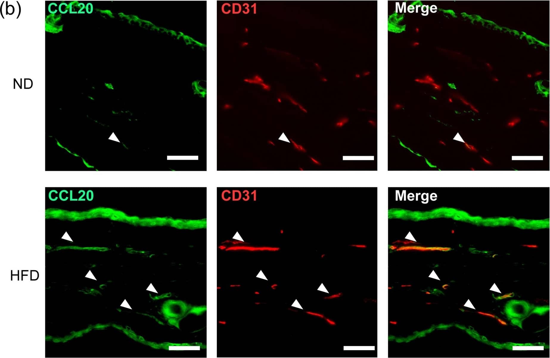
Fatty acid induces Ccl20 expression from dermal blood endothelial cells (BECs) and epidermal keratinocytes. (a) Fold induction of Ccl2, Cxcl16, Ccl20 mRNA in the dermis of ND- or HFD-fed mice in the steady state, as analyzed by qRT-PCR. Results are presented relative to those of ND. The average mRNA expression levels in ND-fed mice are set as 1. (b) Immunohistochemical staining for CCL20 (green) and CD31 (red) in the ear skin of steady state ND and HFD-fed mice. Arrowheads in the panels indicate CCL20+CD31+ cells. Scale bars = 50 μm. (c) Fold induction of Ccl20 mRNA in the steady state epidermis, as analyzed by qRT-PCR. Results are presented relative to those of ND. The average mRNA expression level in ND-fed mice is set as 1. (d) Fold induction of CCL20 mRNA expression in human dermal BECs and epidermal keratinocytes cultured with or without fatty acids for 24 h. Results are presented relative to those of the vehicle-treated group. The average mRNA expression levels in ND-fed mice are set as 1. Pal: palmitic acid, Ste: stearic acid, Ole: oleic acid, Lin: linoleic acid. Results are expressed as the mean ± SEM. p-values were obtained by (a,c) Mann-Whitney-U-test and (d) one way ANOVA. *p ≤ 0.05. Data are pooled from two experiments with four mice (a), two experiments with three mice (c). Data are from one experiment, representative of three independent experiments with three to four mice (b), three experiments performed in triplicate (d). Image collected and cropped by CiteAb from the following open publication (https://pubmed.ncbi.nlm.nih.gov/29074858), licensed under a CC-BY license. Not internally tested by R&D Systems.Detection of Mouse CCL20/MIP-3 alpha by Immunohistochemistry
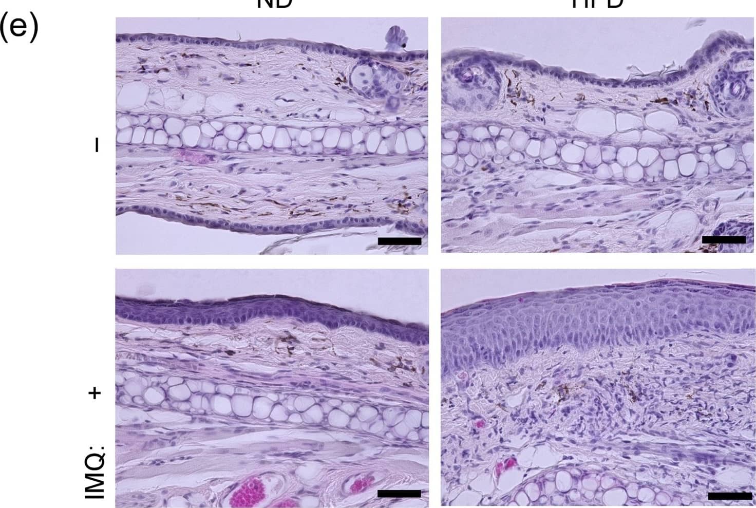
High-fat diet-induced obesity exacerbates imiquimod-induced psoriatic dermatitis. (a) Comparison of body weight between normal diet (ND)- and high fat diet (HFD)-fed mice 10 weeks after feeding. (b–d) Imiquimod (IMQ)-induced psoriatic dermatitis in wild-type (WT) mice fed with either a ND- or HFD-diet. Mice were treated with IMQ for five consecutive days. (b) Representative pictures of psoriatic dermatitis in ear skin. (c) Time course of ear swelling response. (d) Epidermal thickness change with or without IMQ treatment for five days. (e) Representative pictures of H&E-stained sections of the ear skin in ND- and HFD-fed mice with or without IMQ treatment for five days. Samples were collected at 24 h after the last IMQ treatment. Scale bars = 50 μm. Results are expressed as the mean ± SD (c) and SEM (a,d). p-values were obtained by Mann-Whitney-U-test. *p ≤ 0.05. Data are from one experiment, representative of six independent experiments with three to four mice (a), three experiments with three to four mice (b–e). Image collected and cropped by CiteAb from the following open publication (https://pubmed.ncbi.nlm.nih.gov/29074858), licensed under a CC-BY license. Not internally tested by R&D Systems.Applications for Mouse CCL20/MIP-3 alpha Antibody
Western Blot
Sample: Recombinant Mouse CCL20/MIP-3 alpha (Catalog # 760-M3)
Neutralization
Reviewed Applications
Read 1 review rated 4 using AF760 in the following applications:
Formulation, Preparation, and Storage
Purification
Reconstitution
Formulation
Shipping
Stability & Storage
- 12 months from date of receipt, -20 to -70 °C as supplied.
- 1 month, 2 to 8 °C under sterile conditions after reconstitution.
- 6 months, -20 to -70 °C under sterile conditions after reconstitution.
Background: CCL20/MIP-3 alpha
MIP-3 alpha, also known as LARC (Liver and Activation-regulated Chemokine) and Exodus, is one of many novel beta chemokines identified through bioinformatics. Mouse MIP-3 alpha cDNA encodes a 97 amino acid residue precursor protein with a 27 aa residue putative signal peptide that is predicted to be cleaved to form the 70 aa residue mature secreted protein. MIP-3 alpha is distantly related to other beta chemokines (20-28% aa sequence identity). Mouse MIP-3 alpha shares approximately 71% and 63% amino acid sequence homology with rat and human MIP-3 alpha, respectively.
MIP-3 alpha has been shown to be expressed predominantly in lymph nodes, appendix, PBL, fetal liver, fetal lung, and epithelial cells of intestinal tissues. The expression of MIP-3 alpha is strongly up-regulated by inflammatory signals and down-regulated by the anti-inflammatory cytokine IL-10. Synthetic or recombinant MIP-3 alpha has been shown to be chemotactic for lymphocytes and dendritic cells, and inhibits proliferation of myeloid progenitors in colony formation assays. MIP-3 alpha has now been shown to be a unique functional ligand for CCR6 (previously referred to as GPR-CY4, CKR-L3, or STRL22 orphan receptor), a chemokine receptor that is selectively and highly expressed in human dendritic cells derived from CD34+ cord blood precursors.
Alternate Names
Gene Symbol
UniProt
Additional CCL20/MIP-3 alpha Products
Product Documents for Mouse CCL20/MIP-3 alpha Antibody
Product Specific Notices for Mouse CCL20/MIP-3 alpha Antibody
For research use only


