Mouse CD45 Antibody
R&D Systems, part of Bio-Techne | Catalog # MAB114


Key Product Details
Species Reactivity
Validated:
Cited:
Applications
Validated:
Cited:
Label
Antibody Source
Product Specifications
Immunogen
Specificity
Clonality
Host
Isotype
Endotoxin Level
Scientific Data Images for Mouse CD45 Antibody
CD45 in Mouse Splenocytes.
CD45 was detected in immersion fixed mouse splenocytes (positive staining) and Neuro-2A mouse neuroblastoma cell line (negative staining) using Rat Anti-Mouse CD45 Monoclonal Antibody (Catalog # MAB114) at 3 µg/mL for 3 hours at room temperature. Cells were stained using the NorthernLights™ 557-conjugated Anti-Rat IgG Secondary Antibody (red; NL013) and counterstained with DAPI (blue). Specific staining was localized to cell surface. Staining was performed using our protocol for Fluorescent ICC Staining of Non-adherent Cells.CD45 in Mouse Spleen.
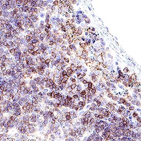
CD45 was detected in immersion fixed paraffin-embedded sections of mouse spleen using Rat Anti-Mouse CD45 Monoclonal Antibody (Catalog # MAB114) at 5 µg/mL for 1 hour at room temperature followed by incubation with the Anti-Rat IgG VisUCyte™ HRP Polymer Antibody (VC005). Before incubation with the primary antibody, tissue was subjected to heat-induced epitope retrieval using Antigen Retrieval Reagent-Basic (CTS013). Tissue was stained using DAB (brown) and counterstained with hematoxylin (blue). Specific staining was localized to cell surface in lymphocytes. Staining was performed using our protocol for IHC Staining with VisUCyte HRP Polymer Detection Reagents.Applications for Mouse CD45 Antibody
Complement-dependent Cytotoxicity
CyTOF-reported
Flow Cytometry
Sample: Mouse splenocytes
Immunocytochemistry
Sample: Immersion fixed mouse splenocytes
Immunohistochemistry
Sample: Immersion fixed paraffin-embedded sections of mouse spleen
Immunoprecipitation
Formulation, Preparation, and Storage
Purification
Reconstitution
Formulation
Shipping
Stability & Storage
- 12 months from date of receipt, -20 to -70 °C as supplied.
- 1 month, 2 to 8 °C under sterile conditions after reconstitution.
- 6 months, -20 to -70 °C under sterile conditions after reconstitution.
Background: CD45
CD45, previously called LCA (leukocyte common antigen), T200, or Ly5 in mouse, is member C of the class 1 (receptor-like) protein tyrosine phosphatase family (PTPRC) (1, 2). It is a variably glycosylated 180‑220 kDa transmembrane protein that is abundantly expressed on all nucleated cells of hematopoietic origin (1‑3). CD45 has several isoforms, expressed according to cell type, developmental stage and antigenic exposure (1‑5). The longest form, CD45RABC (called B220 in mouse), is expressed on B lymphocytes (5). The mouse CD45RABC cDNA encodes 1291 amino acids (aa), including a 23 aa signal sequence, a 541 aa extracellular domain containing the splicing region, a cysteine-rich region and two fibronectin type III domains, a 22 aa transmembrane sequence, and a 705 aa cytoplasmic domain that contains two phosphatase domains, D1 and D2. Only D1 has phosphatase activity. CD45R0 is the shortest form, lacking exons 4, 5 and 6 which encode
aa 30‑169. It is expressed on memory cells, while intermediate sizes are expressed on other T cells (3, 4, 6). CD45 has been best studied in T cells, where it determines T cell receptor signaling thresholds (3, 6‑8). CD45 is moved into or out of the immunological synapse (IS) membrane microdomain depending on the relative influence of interaction with the extracellular galectin lattice or the intracellular actin cytoskeleton (9, 10). Galectin interaction can be fine-tuned by varying usage of the heavily O-glycosylated spliced regions and sialylation of N-linked carbohydrates (4, 9). Within the IS, CD45 dephosphorylates and negatively regulates the src family kinase, LCK (8‑10). In other leukocytes, CD45 influences differentiation and links immunoreceptor signaling with cytokine secretion and cell survival, partially overlapping in function with DEP-1/CD148 (11‑14). CD45 deletion causes in severe immunodeficiency, while point mutations may be associated with autoimmune disorders (6, 7).
References
- Anderson, J.N. et al. (2004) FASEB J. 18:8.
- Streuli, M. et al. (1987) J. Exp. Med. 166:1548.
- Hermiston, M.L. et al. (2003) Annu. Rev. Immunol. 21:107.
- Earl, L.A. and L.G. Baum (2008) Immunol. Cell Biol. 86:608.
- Ralph, S.J. et al. (1987) EMBO J. 6:1251.
- Falahti, R. and D. Leitenberg (2008) J. Immunol. 181:6082.
- Tchilian, E.Z. and P.C.L. Beverley (2006) Trends Immunol. 27:146.
- McNiell, L. et al. (2007) Immunity 27:425.
- Chen, I-J. et al. (2007) J. Biol. Chem. 282:35361.
- Freiberg, B.A. et al. (2002) Nat. Immunol. 3:911.
- Zhu, J.W. et al. (2008) Immunity 28:183.
- Huntington, N.D. et al. (2006) Nat. Immunol. 7:190.
- Hesslein, D.G. et al. (2006) Proc. Natl. Acad. Sci. USA 103:7012.
- Cross, J.L. et al. (2008) J. Immunol. 180:8020.
- Ledbetter, J.A. and L.A. Herzenberg (1979) Immunol. Rev. 47:63.
Long Name
Alternate Names
Gene Symbol
Additional CD45 Products
Product Documents for Mouse CD45 Antibody
Product Specific Notices for Mouse CD45 Antibody
For research use only
