Mouse CXCL9/MIG Antibody
R&D Systems, part of Bio-Techne | Catalog # AF-492-NA


Key Product Details
Species Reactivity
Validated:
Cited:
Applications
Validated:
Cited:
Label
Antibody Source
Product Specifications
Immunogen
Accession # P18340
Specificity
Clonality
Host
Isotype
Endotoxin Level
Scientific Data Images for Mouse CXCL9/MIG Antibody
Chemotaxis Induced by CXCL9/MIG and Neutralization by Mouse CXCL9/MIG Antibody.
Recombinant Mouse CXCL9/MIG (Catalog # 492-MM) chemoattracts the BaF3 mouse pro-B cell line transfected with mouse CXCR3 in a dose-dependent manner (orange line). The amount of cells that migrated through to the lower chemotaxis chamber was measured by Resazurin (Catalog # AR002). Chemotaxis elicited by Recombinant Mouse CXCL9/MIG (1 µg/mL) is neutralized (green line) by increasing concentrations of Goat Anti-Mouse CXCL9/MIG Antigen Affinity-purified Polyclonal Antibody (Catalog # AF-492-NA). The ND50 is typically 6-20 µg/mL.Detection of Mouse CXCL9/MIG by Immunocytochemistry/ Immunofluorescence
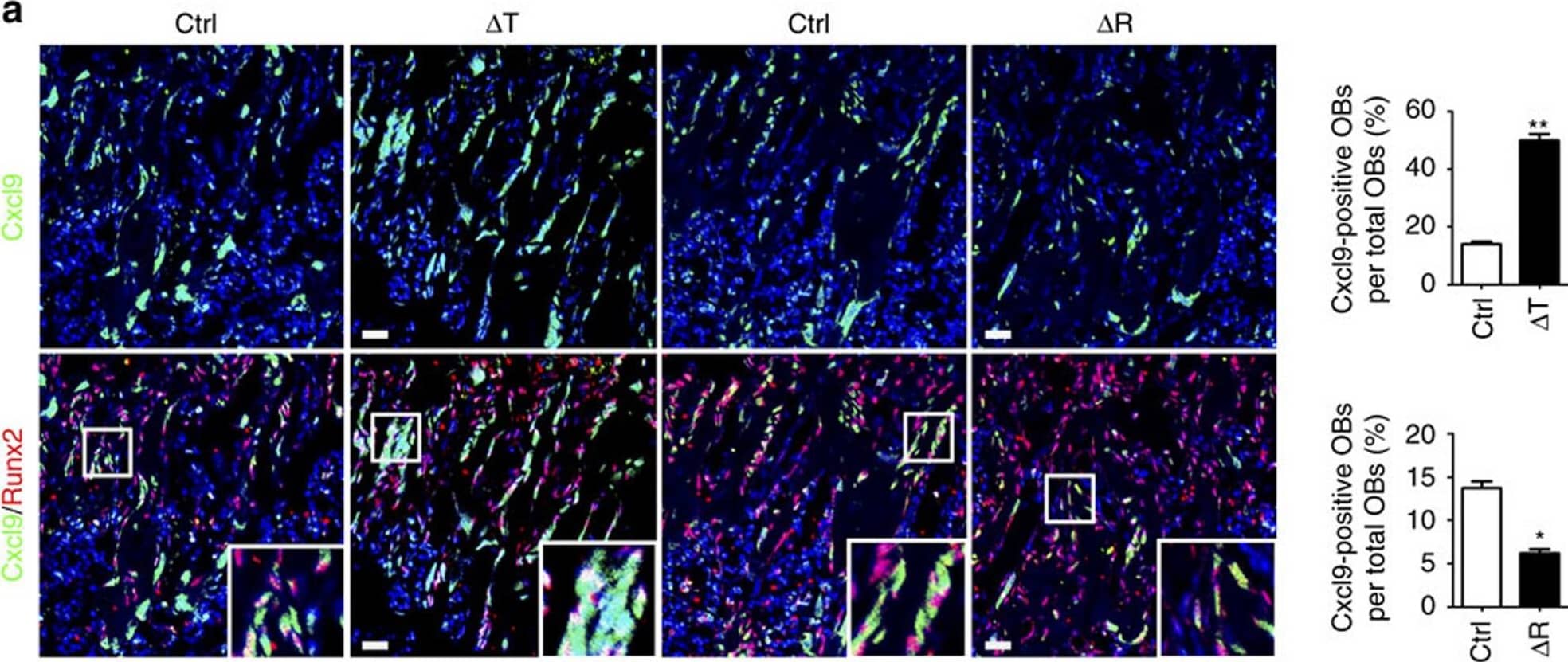
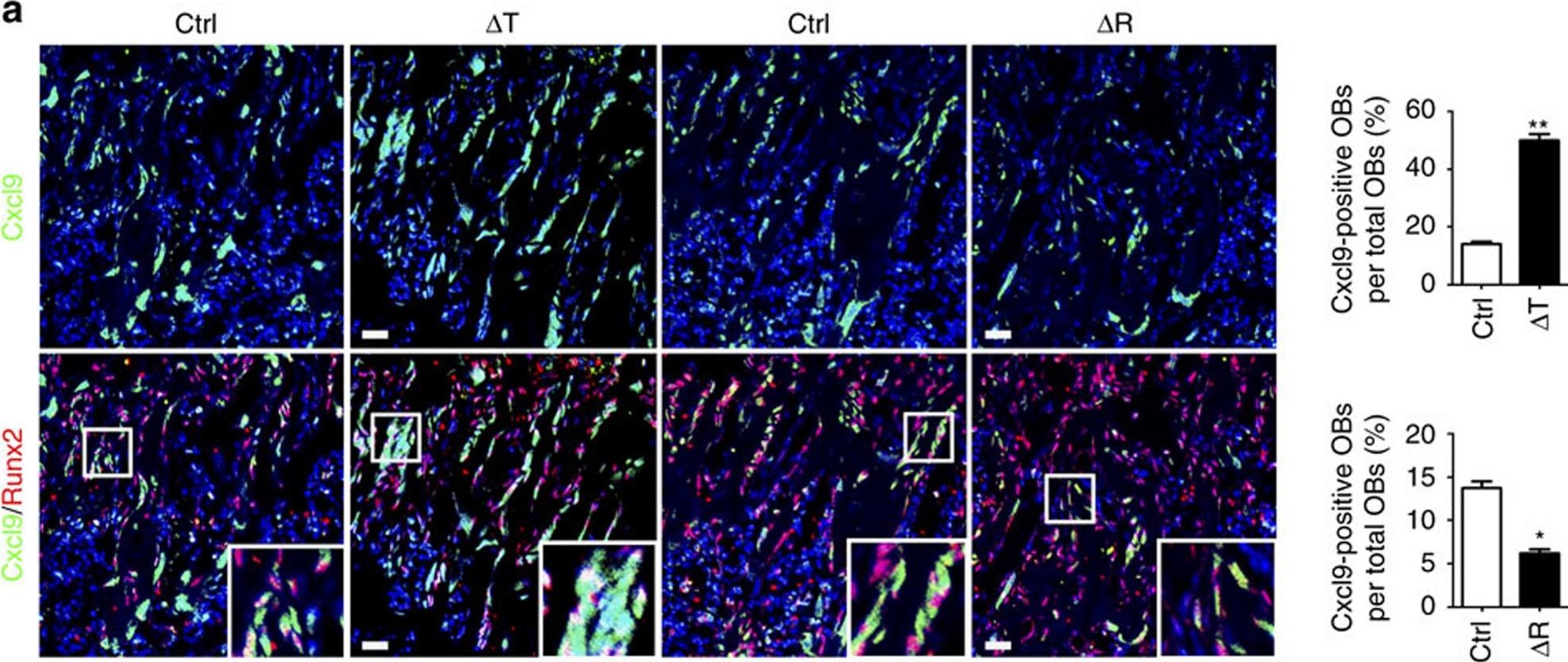
mTORC1 regulates Cxcl9 in osteoblasts.(a) Representative images of in situ hybridization of Cxcl9 mRNA in conjunction with immunostaining of Runx2 in femur sections of 12-week-old male mice bone. Boxed area is enlarged in the bottom right corner. Cxcl9+ osteoblasts out of total osteoblasts were also quantified. Scale bar, 50 μm. n=9 per group. (b) Representative photomicrographs of immunostaining of CXCR3 in CD31+ ECs in bone marrow and quantitative analysis of CXCR3+ ECs out of total ECs in 12-week-old male mice bone. Scale bar, 50 μm. n=9 per group. (c) Representative photomicrographs of immunostaining of CXCR3 in cultured HUVECs. Scale bar, 100 μm. (d) Cxcl9 concentrations assessed by ELISA in bone marrow (BM) and serum. n=5 per group. (e) Quantitative PCR analysis of Cxcl9 mRNA in primary osteoblasts. (f) Western blot of Cxcl9 in primary osteoblasts. (g) Concentrations of Cxcl9 in CM of primary osteoblasts assessed by ELISA. n=5 per group. Data are shown as mean±s.d. *P<0.05, **P<0.01 (Student's t-test). Ctrl, control. Image collected and cropped by CiteAb from the following open publication (https://www.nature.com/articles/ncomms13885), licensed under a CC-BY license. Not internally tested by R&D Systems.Detection of Mouse CXCL9/MIG by Western Blot
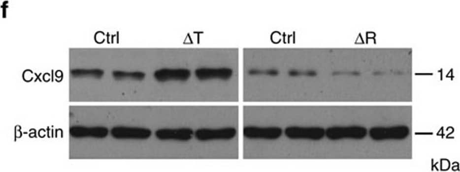
mTORC1 regulates Cxcl9 in osteoblasts.(a) Representative images of in situ hybridization of Cxcl9 mRNA in conjunction with immunostaining of Runx2 in femur sections of 12-week-old male mice bone. Boxed area is enlarged in the bottom right corner. Cxcl9+ osteoblasts out of total osteoblasts were also quantified. Scale bar, 50 μm. n=9 per group. (b) Representative photomicrographs of immunostaining of CXCR3 in CD31+ ECs in bone marrow and quantitative analysis of CXCR3+ ECs out of total ECs in 12-week-old male mice bone. Scale bar, 50 μm. n=9 per group. (c) Representative photomicrographs of immunostaining of CXCR3 in cultured HUVECs. Scale bar, 100 μm. (d) Cxcl9 concentrations assessed by ELISA in bone marrow (BM) and serum. n=5 per group. (e) Quantitative PCR analysis of Cxcl9 mRNA in primary osteoblasts. (f) Western blot of Cxcl9 in primary osteoblasts. (g) Concentrations of Cxcl9 in CM of primary osteoblasts assessed by ELISA. n=5 per group. Data are shown as mean±s.d. *P<0.05, **P<0.01 (Student's t-test). Ctrl, control. Image collected and cropped by CiteAb from the following open publication (https://www.nature.com/articles/ncomms13885), licensed under a CC-BY license. Not internally tested by R&D Systems.Applications for Mouse CXCL9/MIG Antibody
Neutralization
Mouse CXCL9/MIG Sandwich Immunoassay
Formulation, Preparation, and Storage
Purification
Reconstitution
Formulation
Shipping
Stability & Storage
- 12 months from date of receipt, -20 to -70 °C as supplied.
- 1 month, 2 to 8 °C under sterile conditions after reconstitution.
- 6 months, -20 to -70 °C under sterile conditions after reconstitution.
Background: CXCL9/MIG
CXCL9, also known as MIG, is a member of the alpha subfamily of chemokines that lacks the ELR domain, and was initially identified as a lymphokine-activated gene in mouse macrophages. Human CXCL9 was subsequently cloned using mouse CXCL9 cDNA as a probe. The CXCL9 gene is induced in macrophages and in primary glial cells of the central nervous system specifically in response to IFN-gamma. CXCL9 has been shown to be a chemoattractant for activated T-lymphocytes and TIL but not for neutrophils or monocytes. The mouse CXCL9 cDNA encodes a 126 amino acid residue precursor protein with a 21 amino acid residue signal peptide that is cleaved to yield a 105 amino acid residue mature protein. CXCL9 has an extended carboxy-terminus containing greater than 50% basic amino acid residues and is larger than most other chemokines. The carboxy-terminal residues of CXCL9 are prone to proteolytic cleavage resulting in size heterogeneity of natural and recombinant CXCL9. CXCL9 with large carboxy-terminal deletions have been shown to have diminished activity in the calcium flux assay. A chemokine receptor (CXCR3) specific for CXCL9 and IP-10 has been cloned and shown to be highly expressed in IL-2-activated T-lymphocytes.
Alternate Names
Gene Symbol
UniProt
Additional CXCL9/MIG Products
Product Documents for Mouse CXCL9/MIG Antibody
Product Specific Notices for Mouse CXCL9/MIG Antibody
For research use only


