Mouse DNER Antibody
R&D Systems, part of Bio-Techne | Catalog # AF2254


Key Product Details
Species Reactivity
Validated:
Mouse
Cited:
Human, Mouse, Avian - Chicken
Applications
Validated:
Immunohistochemistry, Western Blot
Cited:
Immunocytochemistry, Immunohistochemistry, Immunohistochemistry-Frozen, Immunohistochemistry-Paraffin, Western Blot
Label
Unconjugated
Antibody Source
Polyclonal Goat IgG
Product Specifications
Immunogen
Mouse myeloma cell line NS0-derived recombinant mouse DNER
Ala26-His637
Accession # Q8JZM4
Ala26-His637
Accession # Q8JZM4
Specificity
Detects mouse DNER in direct ELISAs and Western blots. In direct ELISAs and Western blots, approximately 40% cross-reactivity with recombinant human DNER is observed.
Clonality
Polyclonal
Host
Goat
Isotype
IgG
Scientific Data Images for Mouse DNER Antibody
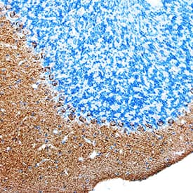
DNER in Mouse Brain.
DNER was detected in perfusion fixed frozen sections of mouse brain (cerebellum) using Goat Anti-Mouse DNER Antigen Affinity-purified Polyclonal Antibody (Catalog # AF2254) at 5 µg/mL overnight at 4 °C. Tissue was stained using the Anti-Goat HRP-DAB Cell & Tissue Staining Kit (brown; Catalog # CTS008) and counterstained with hematoxylin (blue). Specific staining was localized to Purkinje neurons and molecular layer. View our protocol for Chromogenic IHC Staining of Frozen Tissue Sections.Detection of Human DNER by Immunocytochemistry/Immunofluorescence
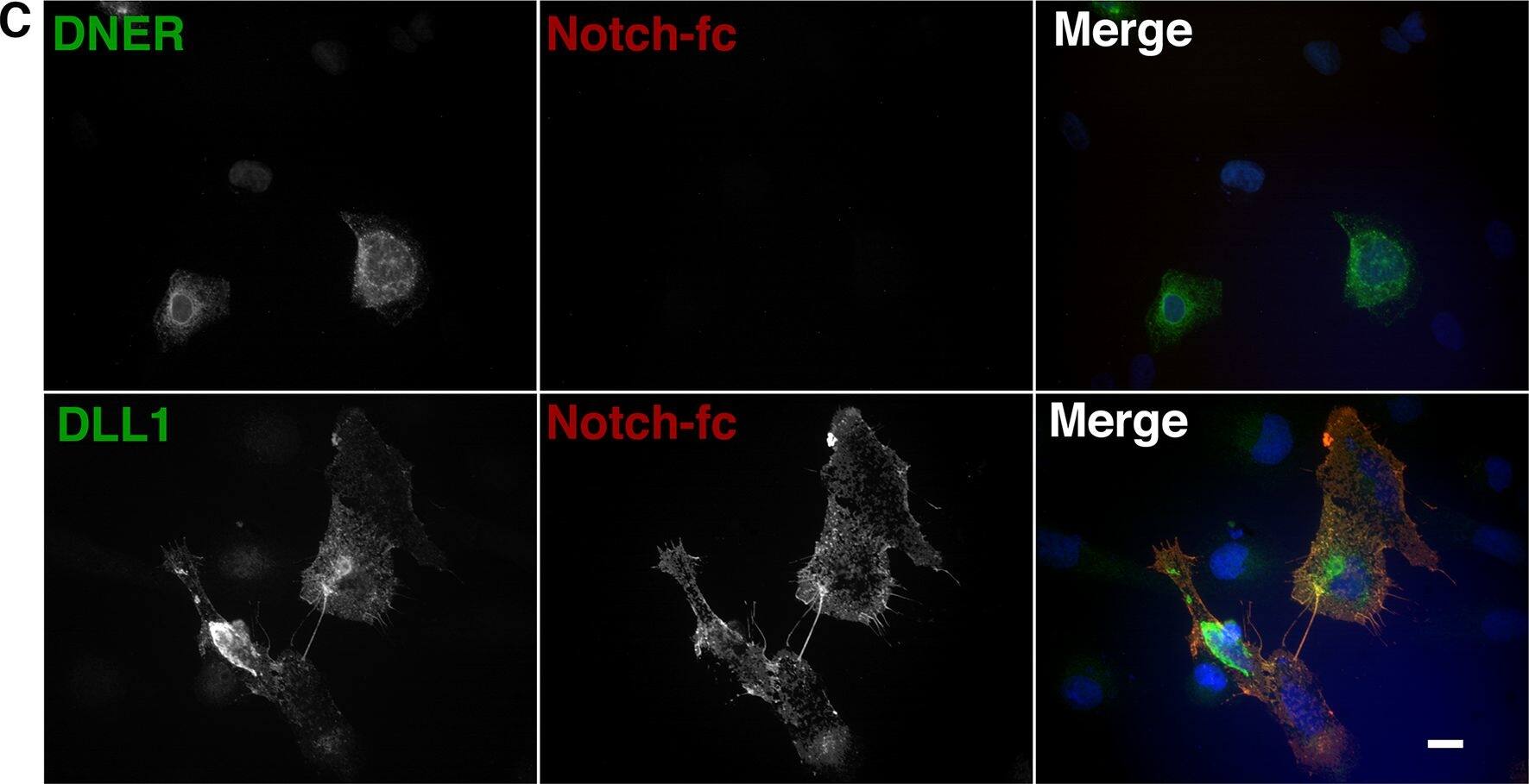
DNER does not activate, function with, or bind to Notch, but known Notch ligand DLL1 does.(A) Pooled luciferase results from 4 separate experiments (normalized to the mean of empty vector in each experiment). U2OS cells were transfected with ligand (DLL1, DNER, or EV), and separately a population of U2OS cells was transfected to express Notch, the control luciferase Renilla, and TP1, a promoter that expresses firefly luciferase when Notch is activated. The two populations were co-cultured 24 hours after transfection (trans configuration), and activity read after an additional 24–48 hours of incubation. (B) C2C12 cells (myoblasts) were incubated with differentiation media (2% horse serum) that either had pre-clustered DLL1-fc (1:1), pre-clustered DNER-fc (1:1), un-clustered DNER-fc, or fc only, all at a ratio of 1:150 in media. Cells were incubated for 72 hours, then fixed, and stained for the presence of myosin heavy chain (MHC) and nuclei. By measuring the percent of total nuclei that were inside of differentiated MHC positive myotubes, fusion indexes were calculated. (C) DNER (top left, green) transfected U2OS cells were not labeled by pre-clustered Notch-fc (top middle, red) but DLL1 (bottom left, green) transfected U2OS cells were labeled by pre-clustered Notch-fc (bottom middle, red). Merged images are shown at far right. Scale 10 μM. **** = p value <0.0001. *** = p value 0.002. ns = not significant. DLL1 = Delta-like 1, a known Notch Ligand, DNER = Delta/Notch-like epidermal growth factor (EGF) related receptor, GSI = gamma-secretase inhibitor, fc only = rabbit anti-human-fc. Image collected and cropped by CiteAb from the following publication (https://dx.plos.org/10.1371/journal.pone.0161157), licensed under a CC-BY license. Not internally tested by R&D Systems.Applications for Mouse DNER Antibody
Application
Recommended Usage
Immunohistochemistry
5-15 µg/mL
Sample: Perfusion fixed frozen sections of mouse brain (cerebellum)
Sample: Perfusion fixed frozen sections of mouse brain (cerebellum)
Western Blot
0.1 µg/mL
Sample: Recombinant Mouse DNER (Catalog # 2254-DN)
Sample: Recombinant Mouse DNER (Catalog # 2254-DN)
Formulation, Preparation, and Storage
Purification
Antigen Affinity-purified
Reconstitution
Reconstitute at 0.2 mg/mL in sterile PBS. For liquid material, refer to CoA for concentration.
Formulation
Lyophilized from a 0.2 μm filtered solution in PBS with Trehalose. *Small pack size (SP) is supplied either lyophilized or as a 0.2 µm filtered solution in PBS.
Shipping
Lyophilized product is shipped at ambient temperature. Liquid small pack size (-SP) is shipped with polar packs. Upon receipt, store immediately at the temperature recommended below.
Stability & Storage
Use a manual defrost freezer and avoid repeated freeze-thaw cycles.
- 12 months from date of receipt, -20 to -70 °C as supplied.
- 1 month, 2 to 8 °C under sterile conditions after reconstitution.
- 6 months, -20 to -70 °C under sterile conditions after reconstitution.
Background: DNER
Long Name
Delta/Notch-like EGF Repeat-containing Transmembrane
Alternate Names
BET, delta and Notch-like epidermal growth factor-related receptor, delta/notch-like EGF repeat containing, delta-notch-like EGF repeat-containing transmembrane, H_NH0150O02.1, UNQ26, WUGSC:H_NH0150O02.1
Gene Symbol
DNER
UniProt
Additional DNER Products
Product Documents for Mouse DNER Antibody
Product Specific Notices for Mouse DNER Antibody
For research use only
Loading...
Loading...
Loading...
Loading...
